The peculiar sensation of hearing your own breathing within your ear can be both disconcerting and fascinating. This phenomenon, known medically as autophony, affects thousands of individuals and represents a complex interplay between the ear’s anatomical structures and various physiological mechanisms. When the delicate balance of your auditory system becomes disrupted, internal sounds that are normally filtered out by your brain suddenly become amplified and noticeable.
Understanding why you can hear yourself breathe in your ear requires exploring the intricate workings of the middle ear, the Eustachian tube system, and the various pathological conditions that can alter normal sound transmission. From simple earwax buildup to complex neurological disorders, numerous factors can contribute to this unsettling auditory experience that impacts quality of life for many sufferers.
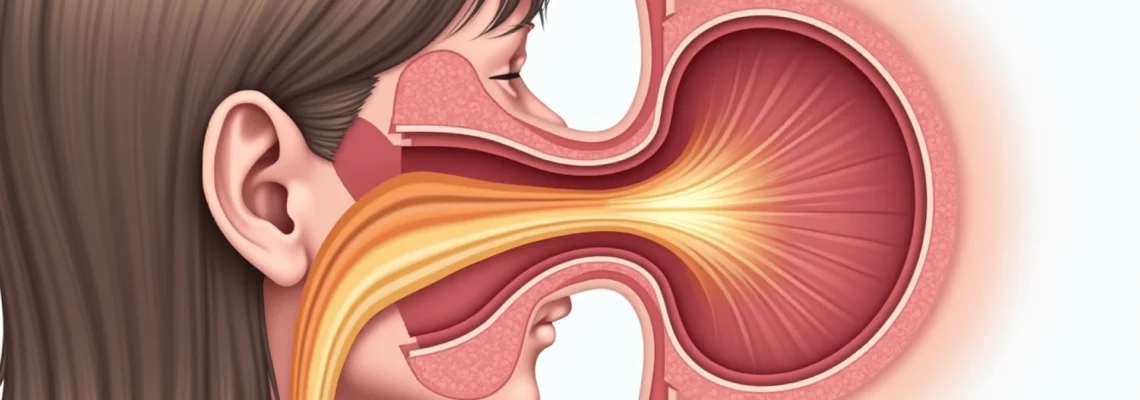
Anatomical structure of the eustachian tube and middle ear cavity
The Eustachian tube serves as a crucial anatomical bridge connecting your middle ear to the nasopharynx, functioning as both a pressure equalisation system and a drainage pathway. This narrow, approximately 35-millimetre-long tube consists of both bony and cartilaginous portions, with the latter comprising roughly two-thirds of its length. The tube’s sophisticated design incorporates muscular control mechanisms that allow it to open and close in response to specific actions like swallowing, yawning, or chewing.
Normal Eustachian tube function relies on the coordinated action of several muscles, particularly the tensor veli palatini and levator veli palatini muscles. These muscles work in harmony to create the opening mechanism that allows air pressure equalisation between the middle ear space and the external atmospheric pressure. When you experience the “popping” sensation during altitude changes, you’re witnessing this remarkable system in action.
Eustachian tube dysfunction and patulous eustachian tube syndrome
Eustachian tube dysfunction occurs when this normally well-coordinated system begins to malfunction, either by remaining closed when it should open or staying open when it should close. The latter condition, known as patulous Eustachian tube syndrome, represents one of the primary causes of hearing your own breathing. In this state, the tube remains abnormally patent, creating an open acoustic pathway that allows respiratory sounds to travel directly from your nasopharynx to your middle ear cavity.
This condition affects approximately 0.3% to 6.6% of the general population, with symptoms ranging from mild intermittent episodes to severe, life-altering manifestations. Patulous Eustachian tube syndrome often develops following significant weight loss, pregnancy, hormonal changes, or certain medications, particularly diuretics that can affect the tissue hydration and muscle tone around the tube.
Middle ear pressure equalisation mechanisms
The middle ear cavity functions as a complex acoustic chamber designed to optimise sound transmission from the external environment to the inner ear. Under normal circumstances, this air-filled space maintains atmospheric pressure through the periodic opening of the Eustachian tube. However, when pressure equalisation mechanisms fail, the resulting pressure differential can significantly amplify internal body sounds, including breathing patterns.
The delicate balance maintained within this system becomes particularly evident during barometric pressure changes or when inflammatory conditions affect the surrounding tissues.
The middle ear’s ability to function as an effective sound transmission system depends entirely on maintaining equal pressure on both sides of the tympanic membrane.
When this equilibrium becomes disrupted, even subtle internal sounds become magnified and perceptible.
Tympanic membrane vibration response to internal sounds
Your eardrum, or tympanic membrane, responds to pressure changes and sound waves with remarkable sensitivity. In cases where the Eustachian tube remains open, respiratory-induced pressure fluctuations cause the tympanic membrane to move synchronously with breathing patterns. This movement creates a direct acoustic coupling between your respiratory system and your auditory perception, resulting in the characteristic sound of hearing your own breathing.
The phenomenon becomes particularly pronounced during deep breathing or physical exertion when respiratory volumes increase. Many individuals report that the sensation intensifies during exercise or when lying in certain positions, reflecting the dynamic nature of this anatomical relationship and its sensitivity to postural and physiological changes.
Mastoid air cell system and acoustic resonance
The mastoid air cell system, located within the temporal bone behind your ear, functions as an extension of the middle ear cavity and contributes significantly to acoustic resonance patterns. This honeycomb-like network of air-filled spaces can amplify certain frequencies, particularly when communication with the middle ear becomes altered due to pathological conditions. Mastoid involvement in autophony often manifests as a hollow, echo-like quality to the perceived internal sounds.
When inflammatory processes affect the mastoid system or when anatomical variations alter normal air circulation patterns, the acoustic properties of the entire middle ear complex can shift dramatically. This can result in enhanced perception of low-frequency sounds, such as breathing, heartbeat, and even eye movements, creating a distressing auditory experience for affected individuals.
Autophony phenomenon and Self-Generated sound transmission
Autophony represents a complex auditory phenomenon where self-generated sounds become abnormally amplified and perceptible. This condition extends beyond simply hearing your breathing to include awareness of your own voice, chewing sounds, heartbeat, and even joint movements. The phenomenon occurs when the normal acoustic filtering mechanisms of the ear become compromised, allowing internal body sounds to bypass the typical dampening systems that prevent such awareness under normal circumstances.
The psychological impact of persistent autophony can be significant, with many individuals reporting decreased quality of life, social withdrawal, and anxiety related to the constant awareness of their internal body sounds. Chronic autophony often leads to hypervigilance regarding ear sensations and can create a cycle of anxiety that potentially worsens the perceived symptoms.
Bone conduction pathways through temporal bone
Sound transmission through bone conduction pathways plays a crucial role in autophony, particularly when normal air conduction mechanisms become altered. The temporal bone serves as an efficient conductor of vibrations from various body structures directly to the inner ear, bypassing the middle ear entirely. This pathway becomes particularly relevant when middle ear pathology exists, as bone-conducted sounds may become relatively enhanced compared to air-conducted sounds.
Understanding bone conduction mechanisms helps explain why certain individuals experience autophony even when their middle ear appears structurally normal. The phenomenon demonstrates how alternative acoustic pathways can become clinically significant when primary transmission routes are compromised, highlighting the sophisticated redundancy built into human auditory systems.
Superior semicircular canal dehiscence syndrome effects
Superior semicircular canal dehiscence syndrome represents a fascinating condition where a small defect in the bone covering the superior semicircular canal creates an abnormal “third window” in the inner ear. This anatomical variant can result in dramatic autophony symptoms, with affected individuals reporting the ability to hear their eye movements, joint creaking, and amplified breathing sounds. The condition affects approximately 0.5% to 0.7% of the population based on temporal bone studies.
Patients with superior canal dehiscence often describe their symptoms as life-changing, with some reporting the ability to hear their own pulse so loudly that it interferes with sleep and concentration.
The diagnosis requires specialised imaging techniques and vestibular testing to confirm the presence of the bony dehiscence and its functional significance.
Respiratory sound amplification in closed ear systems
When the external ear canal becomes occluded by earwax, hearing aid devices, or other obstructions, the acoustic environment within the ear changes dramatically. This closed system can trap and amplify internal sounds, including breathing, creating a phenomenon similar to speaking inside a barrel. The effect becomes particularly noticeable with complete occlusion, where even subtle respiratory movements generate perceivable sound.
Professional earwax removal often provides immediate relief in such cases, demonstrating how simple mechanical obstructions can create complex auditory symptoms. Cerumen impaction remains one of the most treatable causes of autophony, making proper evaluation of ear canal patency essential in any diagnostic workup.
Nasopharyngeal breathing pattern impact on auditory perception
The relationship between breathing patterns and auditory perception becomes particularly evident in individuals with patulous Eustachian tube syndrome. Nasal breathing creates pressure fluctuations that directly transmit to the middle ear through the open tube, while mouth breathing may provide some relief from symptoms. This connection explains why many affected individuals instinctively modify their breathing patterns or seek positions that promote tube closure.
Seasonal allergies and upper respiratory infections can significantly worsen autophony symptoms by altering normal nasopharyngeal anatomy and function. The inflammatory response affects tissue hydration and muscle function around the Eustachian tube, potentially converting a normally functioning tube into a patulous one during acute episodes.
Medical conditions causing enhanced internal sound perception
Numerous medical conditions can contribute to enhanced perception of internal sounds, ranging from common inflammatory disorders to rare neurological conditions. Understanding these underlying pathologies is crucial for developing effective treatment strategies and providing appropriate patient education. The interconnected nature of the auditory system means that disorders affecting seemingly unrelated anatomical structures can manifest as auditory symptoms, including the perception of breathing sounds within the ear.
Systematic evaluation of potential contributing conditions requires comprehensive medical history taking and targeted physical examination. Many conditions present with overlapping symptoms, making differential diagnosis challenging and emphasising the importance of specialised otolaryngological evaluation for persistent cases.
Otosclerosis and stapedial fixation complications
Otosclerosis, characterised by abnormal bone growth around the stapes footplate, can create conductive hearing loss that alters the balance between air-conducted and bone-conducted sound transmission. While primarily causing hearing impairment, the condition can also result in autophony as bone-conducted internal sounds become relatively more prominent compared to diminished air-conducted environmental sounds.
The progressive nature of otosclerosis means that autophony symptoms may develop gradually, often going unrecognised until significant hearing loss occurs. Stapedial fixation affects approximately 0.2% to 0.4% of the population, with women being disproportionately affected, particularly during hormonal changes associated with pregnancy and menopause.
Chronic otitis media with effusion acoustic changes
Chronic otitis media with effusion creates persistent fluid accumulation within the middle ear space, fundamentally altering the acoustic properties of the middle ear system. This fluid can act as a dampening mechanism for external sounds while potentially amplifying internal vibrations transmitted through the skull and surrounding tissues. The condition represents one of the most common causes of conductive hearing loss in children and adults.
The viscosity and composition of middle ear effusion significantly influence the degree of autophony experienced. Thick, mucoid secretions may create different acoustic effects compared to thin, serous fluid, explaining the variability in symptoms reported by affected individuals. Treatment typically focuses on addressing underlying inflammatory conditions and restoring normal Eustachian tube function.
Temporomandibular joint disorder referred symptoms
Temporomandibular joint disorders can produce referred symptoms to the ear region due to the close anatomical proximity and shared nerve pathways. The relationship between jaw function and ear symptoms becomes particularly relevant when considering the muscular connections involved in Eustachian tube function. Dysfunction of the tensor veli palatini muscle, which is innervated by the trigeminal nerve, can affect both jaw function and Eustachian tube opening mechanisms.
TMJ-related autophony often correlates with jaw movement and may fluctuate with chewing patterns or jaw clenching behaviours. The condition demonstrates the interconnected nature of head and neck anatomy and the potential for seemingly unrelated disorders to manifest as auditory symptoms.
Ménière’s disease endolymphatic hydrops effects
Ménière’s disease involves increased pressure within the inner ear’s endolymphatic system, potentially affecting multiple aspects of auditory and vestibular function. While primarily known for causing vertigo and hearing loss, the condition can also result in autophony through alterations in inner ear mechanics and acoustic transmission pathways. The fluctuating nature of symptoms often correlates with changes in endolymphatic pressure.
The unpredictable symptom pattern characteristic of Ménière’s disease can make autophony particularly distressing for affected individuals, as the intensity and character of internal sound perception may vary significantly over time.
Management typically requires a multidisciplinary approach addressing both inner ear pathology and symptom control strategies.
Diagnostic procedures for abnormal breathing sound perception
Comprehensive evaluation of autophony and abnormal breathing sound perception requires systematic assessment utilising multiple diagnostic modalities. The evaluation process typically begins with detailed history taking to characterise the nature, timing, and associated factors related to the symptoms. Clinicians must differentiate between various potential causes while considering the patient’s overall health status, medication use, and recent changes in weight or hormonal status.
Modern diagnostic techniques provide objective measurements that complement clinical observations, enabling more precise diagnosis and treatment planning. The combination of patient-reported symptoms with objective testing results creates a comprehensive picture that guides therapeutic decision-making and helps establish realistic treatment expectations.
Physical examination focuses on otoscopic evaluation of the ear canal and tympanic membrane, assessment of nasal cavity and nasopharynx, and evaluation of Eustachian tube function. Specialised techniques such as nasopharyngeal endoscopy allow direct visualisation of the Eustachian tube opening and assessment of its function during swallowing and other manoeuvres. Tympanometry testing provides objective measurement of middle ear function and can detect abnormal pressure conditions or fluid presence.
Advanced imaging studies may be necessary in cases where structural abnormalities are suspected. High-resolution computed tomography can identify superior canal dehiscence, temporal bone abnormalities, or other anatomical variants contributing to symptoms. Magnetic resonance imaging may be indicated when retrocochlear pathology or inflammatory conditions affecting the cerebellopontine angle are considered in the differential diagnosis.
Audiological assessment plays a crucial role in the diagnostic process, with pure-tone audiometry revealing the degree and configuration of any hearing loss present. Special tests such as acoustic reflex measurements and otoacoustic emissions can provide additional information about middle ear and cochlear function. Vestibular testing may be warranted when superior canal dehiscence syndrome is suspected, as this condition often presents with both auditory and vestibular symptoms.
Treatment protocols and management strategies
Treatment approaches for autophony and breathing sound perception vary significantly based on the underlying cause and symptom severity. Conservative management strategies often provide substantial relief for many individuals, particularly those with mild or intermittent symptoms. The key to successful treatment lies in accurate diagnosis and individualised treatment planning that addresses both the underlying pathology and the patient’s specific symptom profile.
For patulous Eustachian tube syndrome, conservative measures focus on promoting tube closure through postural modifications and nasal treatments. Simple techniques such as lying down with the head dependent can temporarily increase venous congestion in the head and promote tube closure. Nasal saline irrigation and the use of hypertonic saline drops can help maintain adequate tissue hydration around the Eustachian tube opening.
Pharmacological interventions may include nasal decongestants, antihistamines for allergic components, or hormonal treatments in cases related to hormonal fluctuations. However, caution must be exercised with decongestant use, as overuse can potentially worsen Eustachian tube function. Some patients benefit from specialty compounds such as chlorobutanol or benzyl alcohol nasal drops, which may help promote tube closure through local tissue effects.
Surgical interventions are reserved for severe cases that fail to respond to conservative management. Options include tympanic membrane procedures such as myringotomy with tube placement, which can equalise middle ear pressure and reduce autophony symptoms. More complex procedures may involve Eustachian tube reconstruction or plugging procedures, though these carry higher risks and variable success rates.
For conditions such as superior canal dehiscence syndrome, surgical repair through middle cranial fossa or mastoid approaches can provide definitive treatment. The choice of surgical approach depends on factors such as the size and location of the dehiscence, patient age and overall health, and the severity of symptoms. Success rates for properly selected patients exceed 90% for resolution of autophony symptoms.
Preventive measures and lifestyle modifications for ear health
Preventive strategies for maintaining optimal ear health and reducing the risk of developing autophony focus on supporting normal Eustachian tube function and avoiding factors that can disrupt the delicate balance of middle ear
mechanics. These strategies encompass both general health practices and specific measures designed to support optimal auditory system function throughout life.
Weight management plays a crucial role in preventing patulous Eustachian tube syndrome, as rapid weight loss represents one of the most significant risk factors for developing this condition. Maintaining stable body weight through balanced nutrition and regular exercise helps preserve the fatty tissue and muscle tone necessary for proper Eustachian tube function. Individuals who must undergo significant weight loss should work closely with healthcare providers to monitor for early signs of Eustachian tube dysfunction.
Proper hydration supports the maintenance of healthy mucous membranes throughout the upper respiratory tract and middle ear system. Adequate fluid intake helps maintain optimal tissue hydration around the Eustachian tube opening, preventing the tissue changes that can contribute to abnormal tube function. The general recommendation of 8-10 glasses of water daily becomes particularly important for individuals with a history of upper respiratory infections or allergic conditions.
Environmental factors significantly influence ear health, with particular attention needed for allergen exposure and air quality. Regular cleaning of living and working spaces to reduce dust, mould, and other allergens can help prevent the inflammatory conditions that commonly trigger Eustachian tube dysfunction. Indoor air quality management through proper ventilation, humidity control, and air filtration systems creates an environment that supports healthy respiratory and auditory function.
Medication awareness becomes essential for preventing drug-induced autophony, particularly with diuretics and other medications that can affect tissue hydration and electrolyte balance. Patients prescribed these medications should be counselled about potential auditory side effects and encouraged to report any new ear-related symptoms promptly. Regular monitoring allows for early intervention before symptoms become severe or persistent.
Prevention of autophony often involves addressing seemingly unrelated health factors, demonstrating how interconnected body systems require comprehensive health management for optimal function.
Stress management techniques contribute to overall ear health by reducing the muscle tension and inflammatory responses that can affect Eustachian tube function. Chronic stress has been associated with increased risk of upper respiratory infections and allergic responses, both of which can trigger auditory symptoms. Regular exercise, adequate sleep, and stress reduction practices create a foundation for maintaining healthy auditory system function throughout life.
Regular hearing health assessments should be integrated into routine healthcare, particularly for individuals over 50 or those with risk factors for hearing disorders. Early detection of hearing changes or ear-related symptoms allows for prompt intervention before conditions progress to more severe stages. Professional ear cleaning and examination can identify potential problems such as earwax accumulation or early signs of infection before they cause significant symptoms.
Understanding when to seek professional help empowers individuals to take appropriate action when symptoms develop. Persistent autophony lasting more than a few days, symptoms that worsen over time, or autophony associated with hearing loss, dizziness, or pain warrant prompt medical evaluation. Early intervention often provides better treatment outcomes and can prevent the development of chronic symptoms that significantly impact quality of life.