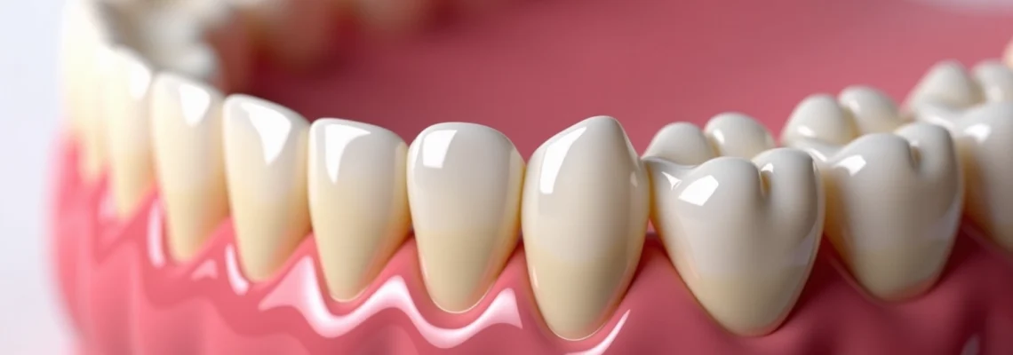
Have you ever noticed that the edges of your teeth appear translucent or clear when you smile in the mirror? This phenomenon, known as incisal edge transparency, is actually quite common and can occur for various reasons ranging from natural tooth development to pathological conditions. The translucent appearance at the bottom of your teeth is primarily related to the unique structure and thickness of dental enamel in this region, combined with the way light interacts with your tooth surfaces.
Understanding why teeth develop this transparent quality is crucial for both dental professionals and patients alike. Enamel translucency can indicate normal physiological processes or signal underlying dental health concerns that require professional attention. The complexity of tooth structure, particularly the intricate relationship between enamel thickness, mineral content, and light transmission properties, plays a fundamental role in determining the optical characteristics of your teeth.
Dental enamel structure and transparency mechanisms
The transparency observed at the bottom edges of teeth stems from the sophisticated architectural design of dental enamel. This remarkable biological material consists of highly organised crystalline structures that interact with light in unique ways, creating the optical properties we observe clinically. The transparency phenomenon occurs due to the specific arrangement of enamel components and their varying densities throughout different regions of the tooth crown.
Hydroxyapatite crystal composition in incisal edges
Dental enamel comprises approximately 96% hydroxyapatite crystals, which are arranged in a highly organised hexagonal pattern. At the incisal edges, these crystals are more densely packed and aligned, creating pathways for light transmission. The crystalline structure at the tooth edges contains fewer organic materials and more mineral content compared to other enamel regions, contributing to increased translucency. This mineral-rich composition allows light to pass through more readily, creating the characteristic transparent appearance that many individuals observe.
Enamel rod orientation and light refraction properties
Enamel rods, also known as prisms, extend from the dentine-enamel junction to the tooth surface in a complex three-dimensional pattern. Near the incisal edges, these rods run more parallel to the tooth surface, creating optimal conditions for light transmission. The rod orientation significantly influences how light travels through the enamel structure, with parallel arrangements facilitating greater translucency. This architectural feature explains why the edges of teeth naturally appear more transparent than the central portions, where enamel rods follow more complex pathways.
Developmental enamel thickness variations
During tooth development, enamel formation occurs in stages, with varying thickness patterns across the crown surface. The incisal edges typically develop with thinner enamel layers compared to the cervical regions, creating natural variations in opacity. Amelogenesis , the process of enamel formation, produces these thickness gradients as part of normal tooth morphology. These developmental variations contribute to the natural translucency observed at tooth edges and represent normal anatomical features rather than pathological conditions.
Dentine-enamel junction translucency effects
The interface between enamel and underlying dentine plays a crucial role in tooth transparency. At the incisal edges, this junction may be more pronounced due to the convergence of enamel and dentine layers. The scalloped morphology of the dentine-enamel junction creates micro-spaces that can affect light transmission properties. Additionally, the yellowish colour of dentine becomes more apparent through thinner enamel sections, contributing to the overall optical characteristics of the tooth edge.
Physiological causes of incisal edge transparency
Natural physiological processes throughout life can contribute to increased transparency at the bottom of teeth. These changes occur as part of normal aging, functional wear, and individual genetic variations that affect enamel development and maintenance. Understanding these physiological factors helps distinguish between normal age-related changes and pathological conditions requiring intervention.
Natural enamel thinning through masticatory function
Daily masticatory activities, including chewing, biting, and speaking, gradually wear away microscopic amounts of enamel from the incisal edges. This physiological attrition is a normal part of tooth function but can progressively increase transparency over time. The mechanical forces generated during normal function create micro-abrasions that slowly reduce enamel thickness at the contact points. Modern dietary patterns, particularly the consumption of processed foods, may accelerate this natural wearing process compared to ancestral diets.
Age-related enamel wear patterns and attrition
As individuals age, cumulative wear patterns develop on tooth surfaces, particularly affecting the incisal edges where forces concentrate during function. Age-related changes in enamel structure include mineral loss, micro-crack formation, and gradual thinning of the protective layer. These changes typically begin in the third decade of life and progress steadily, with transparency becoming more noticeable after age 40. The wear patterns reflect individual functional habits, dietary preferences, and oral parafunctional activities accumulated over years of use.
Bruxism-induced enamel erosion at crown margins
Teeth grinding and clenching, known as bruxism, creates excessive mechanical stress on tooth structures, particularly affecting the incisal edges. The repetitive grinding motions during sleep or stress-related clenching generate forces that can exceed normal physiological limits. Bruxism-related wear accelerates enamel loss at the crown margins, creating characteristic flattened surfaces with increased transparency. This condition affects approximately 8-15% of adults and can significantly impact tooth appearance and structural integrity.
Genetic predisposition to thin enamel formation
Genetic factors influence enamel thickness, mineral content, and structural organisation, with some individuals inheriting predispositions to thinner enamel formation. Hereditary enamel defects can manifest as generalised thinning or localised weak areas that appear more translucent. Genetic variations in amelogenin proteins, which guide enamel formation, can result in structural abnormalities that affect optical properties. Family history of thin enamel or early tooth wear may indicate genetic susceptibility to transparency development.
Pathological conditions affecting enamel opacity
Several pathological conditions can accelerate enamel loss and increase tooth transparency beyond normal physiological limits. Erosive tooth wear represents one of the most significant challenges in modern dentistry, with acid-related enamel loss affecting substantial portions of the population. Gastroesophageal reflux disease (GERD) exposes teeth to stomach acids with pH levels below 2.0, creating severe erosive conditions that rapidly dissolve enamel minerals. This internal acid exposure primarily affects lingual surfaces but can extend to incisal edges in severe cases.
Dietary acid exposure from frequent consumption of carbonated beverages, citrus fruits, and sports drinks creates an acidic oral environment that promotes enamel demineralisation. The critical pH threshold for enamel dissolution is approximately 5.5, and many common beverages fall well below this level. Bulimia nervosa presents particularly severe challenges, with repeated vomiting episodes exposing teeth to stomach acids and creating characteristic erosion patterns on lingual surfaces that extend to incisal edges.
Amelogenesis imperfecta represents a group of inherited disorders affecting enamel formation, resulting in teeth with abnormal structure, thickness, and appearance. These conditions can cause generalised enamel hypoplasia, hypomaturation, or hypocalcification, all of which contribute to increased translucency. Celiac disease has been associated with enamel defects, particularly affecting permanent teeth development during childhood. The autoimmune response in celiac disease can interfere with normal amelogenesis, resulting in characteristic enamel abnormalities including increased transparency.
Fluorosis, caused by excessive fluoride exposure during tooth development, can paradoxically increase enamel translucency in severe cases. While mild fluorosis strengthens enamel, excessive exposure creates structural abnormalities that affect optical properties. Medication-induced enamel changes from tetracycline antibiotics, chemotherapy agents, or chronic use of acidic medications can also contribute to structural alterations affecting transparency.
Clinical assessment techniques for enamel translucency
Accurate assessment of enamel translucency requires sophisticated diagnostic approaches that combine visual examination with advanced imaging techniques. Modern dental practice utilises multiple assessment methods to evaluate the extent and progression of transparency changes, enabling precise treatment planning and monitoring.
Transillumination diagnostic methods using Fibre-Optic light
Transillumination represents the gold standard for assessing enamel translucency and internal tooth structure. Fibre-optic light sources positioned at specific angles reveal the extent of transparency and help identify areas of enamel thinning. LED transillumination devices provide consistent light output and enable standardised assessment protocols. The technique involves positioning the light source palatally or lingually while observing from the facial aspect, creating distinctive patterns that indicate enamel thickness variations.
Digital photography documentation with polarised filters
High-resolution digital photography using polarised filters eliminates surface reflections and enhances visualisation of enamel translucency patterns. Cross-polarisation techniques involve placing filters over both the light source and camera lens to eliminate surface glare. Standardised photographic protocols ensure consistent documentation for comparison over time and facilitate communication between practitioners. These imaging techniques prove particularly valuable for monitoring progression and evaluating treatment outcomes.
VITA classical shade guide translucency matching
The VITA Classical shade guide includes translucency parameters that help standardise assessment and communication regarding enamel opacity changes. Professional shade matching considers both colour and translucency characteristics to achieve optimal aesthetic outcomes. Spectrophotometric analysis provides objective measurements of light transmission properties, eliminating subjective variations in visual assessment. These standardised approaches ensure consistent evaluation across different practitioners and clinical settings.
Restorative treatment options for transparent incisal edges
Contemporary restorative dentistry offers multiple approaches for addressing transparent incisal edges, ranging from conservative enamel preservation techniques to comprehensive aesthetic reconstruction. Treatment selection depends on the extent of transparency, underlying causes, functional requirements, and patient aesthetic expectations.
Composite resin layering with IPS empress direct materials
Advanced composite resin systems like IPS Empress Direct offer excellent optical properties that can effectively mask transparent areas while preserving natural tooth structure. The layering technique involves applying multiple thin increments with varying opacity levels to recreate natural enamel characteristics. Polychromatic layering utilises different composite shades and translucencies to achieve seamless integration with existing tooth structure. This conservative approach preserves maximum natural enamel while addressing aesthetic concerns effectively.
Porcelain veneer applications using feldspathic ceramics
Feldspathic ceramic veneers provide superior aesthetic results for severe transparency cases requiring comprehensive coverage. These ultra-thin restorations, typically 0.3-0.5mm thick, can effectively mask underlying transparency while maintaining natural appearance. Preparation design for transparent edges requires careful consideration of reduction depths to ensure adequate ceramic thickness for opacity control. Modern ceramic systems offer excellent light transmission properties that mimic natural enamel behaviour.
Enamel microabrasion techniques with 18% hydrochloric acid
Enamel microabrasion using controlled acid etching can improve the appearance of superficial transparency by removing the outermost enamel layer. The procedure involves applying 18% hydrochloric acid combined with pumice to create controlled enamel removal. Microabrasion protocols require precise application techniques and careful monitoring to prevent excessive enamel loss. This conservative approach works best for mild transparency cases without significant structural compromise.
Bonding agent selection for optimal light transmission
The choice of bonding agents significantly affects the optical properties of composite restorations on transparent teeth. Modern adhesive systems must provide strong bonding while maintaining appropriate light transmission characteristics. Universal bonding agents offer versatility for different substrates and can be used in total-etch or self-etch modes depending on clinical requirements. Proper selection and application of bonding systems ensure long-term success and optimal aesthetic integration with natural tooth structure.