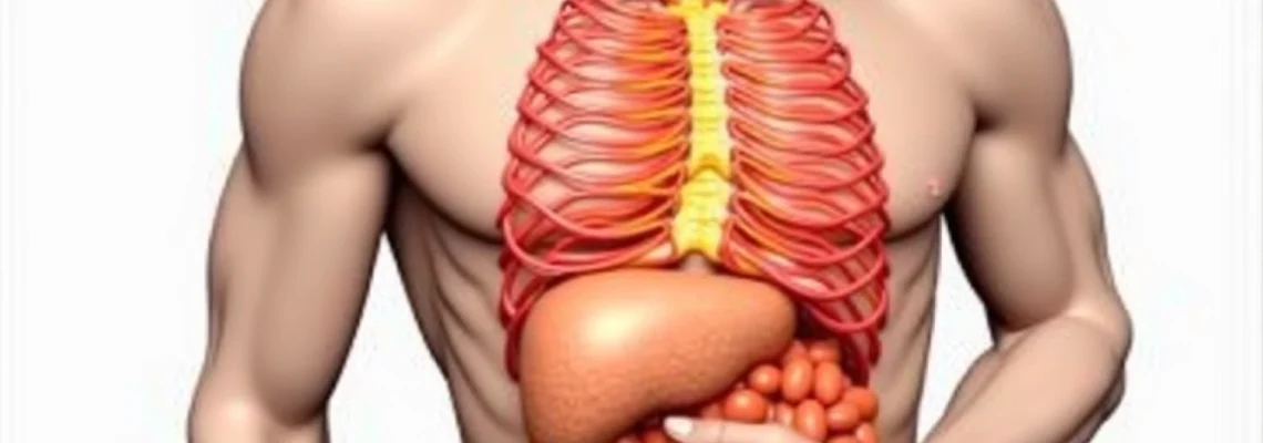
Sharp pain beneath the right rib cage can be an alarming experience that affects millions of people worldwide. This type of discomfort, medically termed right upper quadrant (RUQ) pain, encompasses a broad spectrum of potential causes ranging from benign muscle strains to serious hepatobiliary disorders. The right subcostal region houses several vital organs, including the liver, gallbladder, right kidney, and portions of the intestines, making accurate diagnosis crucial for appropriate treatment.
Understanding the underlying mechanisms of right subcostal pain requires careful consideration of anatomical relationships and physiological processes. The complexity of this region means that pain can originate from hepatic capsule distension, biliary obstruction, musculoskeletal inflammation, or even referred pain from pulmonary pathologies. Early recognition of concerning symptoms can significantly impact treatment outcomes and prevent potentially life-threatening complications.
Hepatobiliary disorders causing right subcostal pain
The hepatobiliary system represents the most common source of right subcostal pain, with gallbladder-related conditions accounting for approximately 65% of all RUQ complaints. These disorders often present with characteristic pain patterns that can help clinicians differentiate between various pathological processes affecting the liver, gallbladder, and biliary tree.
Acute cholecystitis and gallbladder inflammation
Acute cholecystitis stands as the predominant cause of severe right subcostal pain, affecting nearly 20 million Americans annually. This condition typically develops when gallstones obstruct the cystic duct, leading to gallbladder distension and subsequent inflammatory response. The resulting pain is characteristically sharp and persistent , often radiating to the right shoulder blade or scapular region.
The pathophysiology involves increased intraluminal pressure within the gallbladder, causing ischaemia of the gallbladder wall and triggering an inflammatory cascade. Patients frequently describe the pain as reaching peak intensity within hours of onset, accompanied by nausea, vomiting, and low-grade fever. Murphy’s sign, characterised by inspiratory arrest during deep palpation of the right subcostal margin, remains a reliable clinical indicator of acute cholecystitis.
Clinical studies demonstrate that delayed treatment of acute cholecystitis increases the risk of complications, including gallbladder perforation and sepsis, by up to 15% within the first 72 hours of symptom onset.
Choledocholithiasis and common bile duct obstruction
Choledocholithiasis, the presence of gallstones within the common bile duct, creates a distinct pattern of right subcostal pain that differs from simple cholecystitis. This condition affects approximately 10-15% of patients with gallstone disease and can lead to serious complications if left untreated. The pain associated with bile duct obstruction tends to be more severe and cramping in nature, reflecting the smooth muscle spasms as the biliary tree attempts to overcome the obstruction.
The classic presentation includes Charcot’s triad: right upper quadrant pain, jaundice, and fever. However, complete triad presentation occurs in only 50-75% of cases, making diagnosis challenging. Jaundice typically develops when bilirubin levels exceed 2.5 mg/dL, manifesting as yellowing of the sclera and skin. Laboratory investigations reveal elevated alkaline phosphatase, gamma-glutamyl transferase, and conjugated bilirubin levels, supporting the diagnosis of biliary obstruction.
Hepatitis and liver capsule distension
Hepatitis, whether viral, autoimmune, or drug-induced, can cause significant right subcostal discomfort through liver capsule stretching. The liver capsule contains numerous pain-sensitive nerve fibres, and any condition causing hepatomegaly will trigger nociceptive responses. Unlike gallbladder pain, hepatitis-related discomfort tends to be more constant and aching rather than sharp or cramping.
Acute viral hepatitis affects approximately 4.4 million Americans annually, with hepatitis A, B, and C representing the most common causes. The pain pattern typically correlates with the degree of hepatocellular inflammation and liver enlargement. Patients may also experience systemic symptoms including fatigue, anorexia, and low-grade fever. Drug-induced hepatitis from paracetamol overdose or other hepatotoxic medications can present similarly but often with more rapid onset and potentially greater severity.
Cholangitis and biliary tree infections
Ascending cholangitis represents a medical emergency characterised by bacterial infection of the biliary tree, typically occurring secondary to biliary obstruction. This condition affects approximately 1-3% of patients with choledocholithiasis but carries significant morbidity and mortality risks if not promptly treated. The pain associated with cholangitis is typically severe and constant, often described as deep and aching.
Reynolds’ pentad, which includes Charcot’s triad plus hypotension and altered mental status, indicates severe cholangitis requiring immediate intervention. The pathogenic organisms commonly include Escherichia coli, Klebsiella species, and Enterococcus. Early recognition and treatment with broad-spectrum antibiotics and biliary drainage can significantly improve outcomes, with mortality rates decreasing from 50% to less than 10% with appropriate management.
Musculoskeletal aetiologies of right costal margin pain
Musculoskeletal causes represent the second most common source of right subcostal pain, accounting for approximately 25-30% of cases. These conditions often present with movement-related pain patterns that can help differentiate them from visceral causes. The complexity of the thoracic musculature and innervation patterns means that seemingly minor injuries can produce significant discomfort.
Intercostal neuralgia and nerve entrapment syndromes
Intercostal neuralgia affects the thoracic spinal nerves T1-T12, causing sharp, shooting pain that follows the course of the affected nerve. This condition can result from various causes, including post-herpetic neuralgia, thoracic surgery, or direct trauma. The pain typically presents as a band-like sensation extending from the spine around to the anterior chest wall, often intensifying with deep inspiration or movement.
Nerve entrapment syndromes, particularly involving the lateral cutaneous branches of the intercostal nerves, can create localised areas of intense pain beneath the right rib cage. These conditions often develop following repetitive activities or sustained positions that compress neural structures. Diagnostic nerve blocks can provide both therapeutic relief and confirmatory evidence of nerve involvement. Treatment approaches include neuropathic pain medications, local anaesthetic injections, and physical therapy modalities.
Costochondritis and tietze syndrome manifestations
Costochondritis involves inflammation of the costochondral junctions, creating localised tenderness and pain that can mimic cardiac or pulmonary pathology. This condition affects multiple age groups but shows particular prevalence in individuals aged 20-40 years. The pain characteristically worsens with chest wall movement, deep breathing, or direct palpation of the affected costal cartilages.
Tietze syndrome represents a specific variant characterised by visible swelling and tenderness of the costochondral or costosternal junctions, typically affecting the second and third ribs. Unlike costochondritis, Tietze syndrome presents with obvious inflammatory signs and tends to affect younger patients. Anti-inflammatory medications and physical therapy techniques focusing on chest wall mobility provide the primary treatment approach. Heat therapy and gentle stretching exercises can also contribute to symptom resolution.
Rib fractures and thoracic cage trauma
Rib fractures represent a significant cause of acute right subcostal pain, particularly following trauma or in elderly patients with osteoporosis. The lower ribs (8-12) are more susceptible to injury due to their reduced protection from overlying musculature. Fractures typically cause severe, sharp pain that intensifies dramatically with respiratory movements, coughing, or positional changes.
Stress fractures can develop in athletes or individuals with repetitive overhead activities, creating chronic pain that may initially be subtle but progressively worsens. The healing process typically requires 6-8 weeks, during which pain management and breathing exercises remain crucial to prevent complications such as pneumonia. Flail chest , involving multiple rib fractures, represents a medical emergency requiring immediate stabilisation and respiratory support.
Myofascial pain syndrome in the right thoracic region
Myofascial pain syndrome affects the fascial layers and musculature surrounding the thoracic cage, creating trigger points that can refer pain to the subcostal region. This condition often develops following repetitive strain, poor posture, or psychological stress. The pain pattern typically involves deep, aching sensations with associated muscle stiffness and reduced range of motion.
Trigger points in the serratus anterior, latissimus dorsi, or external oblique muscles can create referred pain patterns that mimic visceral pathology. Manual therapy techniques, including trigger point release and myofascial stretching, provide effective treatment approaches. Postural correction and ergonomic modifications often play crucial roles in preventing recurrence and maintaining long-term symptom relief.
Pulmonary pathologies presenting as right subcostal pain
Respiratory conditions can frequently manifest as right subcostal pain due to the anatomical relationships between the lungs, pleura, and diaphragm. The lower lobe of the right lung extends to the level of the sixth rib anteriorly, making pulmonary pathology a potential cause of subcostal discomfort. Understanding these relationships helps clinicians recognise when apparently abdominal pain may actually originate from thoracic structures.
Pleuritic pain and pleural effusion
Pleuritic pain results from inflammation of the parietal pleura, which contains abundant pain-sensitive nerve fibres supplied by the intercostal and phrenic nerves. This condition creates characteristic sharp, stabbing pain that intensifies dramatically with inspiration, coughing, or movement. The pain typically has a knife-like quality and can cause patients to adopt shallow breathing patterns to minimise discomfort.
Pleural effusion, the accumulation of fluid within the pleural space, can cause a dull, aching sensation in the subcostal region as the lung becomes compressed and the pleural surfaces stretch. Large effusions may create a sensation of fullness or pressure rather than sharp pain. Diagnostic thoracentesis not only provides tissue samples for analysis but often yields immediate symptomatic relief. The underlying cause, whether infectious, malignant, or inflammatory, determines the specific treatment approach.
Pneumonia with diaphragmatic irritation
Lower lobe pneumonia can present with pain referred to the subcostal region through diaphragmatic irritation. The phrenic nerve, which supplies both the diaphragm and provides sensory innervation to the peritoneum, can create confusing pain patterns that mimic abdominal pathology. This phenomenon occurs in approximately 20-30% of lower lobe pneumonia cases and can delay appropriate diagnosis.
The inflammatory process within the lung parenchyma stimulates pleural pain receptors, while diaphragmatic involvement creates referred pain to the ipsilateral shoulder and subcostal region. Elderly patients and those with compromised immune systems may present with subtle symptoms, making clinical recognition challenging. High-resolution computed tomography provides superior diagnostic accuracy compared to standard chest radiographs, particularly in cases with atypical presentations.
Recent studies indicate that lower lobe pneumonia with diaphragmatic involvement accounts for up to 15% of cases initially misdiagnosed as biliary or hepatic pathology in emergency department settings.
Pulmonary embolism and Right-Sided chest pain
Pulmonary embolism (PE) represents a potentially life-threatening condition that can present with right subcostal pain, particularly when involving the lower lobe pulmonary vessels. The pain associated with PE often has a pleuritic component but can also manifest as a constant, dull ache. Risk factors include prolonged immobilisation, recent surgery, malignancy, and hypercoagulable states.
The Wells score provides a validated clinical prediction tool for assessing PE probability, incorporating factors such as clinical signs of deep vein thrombosis, alternative diagnoses, and risk factors. D-dimer levels, while sensitive, lack specificity and must be interpreted within the appropriate clinical context. CT pulmonary angiography remains the gold standard for diagnosis, providing detailed visualisation of the pulmonary vasculature and identification of embolic material.
Pneumothorax and pleural space complications
Pneumothorax, whether spontaneous or traumatic, can cause sudden onset right subcostal pain accompanied by dyspnoea and chest tightness. Primary spontaneous pneumothorax typically affects tall, thin young adults, while secondary pneumothorax occurs in patients with underlying lung disease. The pain pattern often begins sharply and may diminish as the lung collapses and pleural surfaces separate.
Tension pneumothorax represents a medical emergency requiring immediate needle decompression followed by tube thoracostomy. The progressive accumulation of air under pressure can compromise venous return and cardiac output, leading to haemodynamic instability. Clinical signs include tracheal deviation, absent breath sounds, and jugular venous distension. Recognition and treatment within minutes can be life-saving, emphasising the importance of maintaining high clinical suspicion in appropriate scenarios.
Gastrointestinal conditions mimicking right subcostal discomfort
Various gastrointestinal pathologies can present with pain in the right subcostal region, creating diagnostic challenges for healthcare providers. The complex innervation patterns of the gastrointestinal tract, combined with the phenomenon of referred pain, mean that conditions affecting distant organs can manifest as localised subcostal discomfort. Understanding these pain referral patterns is crucial for accurate diagnosis and appropriate management.
Peptic ulcer disease, particularly duodenal ulcers, can cause pain that radiates to the right subcostal area, especially during periods of gastric acid hypersecretion. The pain typically demonstrates a characteristic relationship with meals, often improving with food intake but recurring several hours later as gastric emptying occurs. Helicobacter pylori infection remains the predominant cause of peptic ulceration, affecting approximately 50% of the global population and requiring specific antibiotic therapy for eradication.
Gastroesophageal reflux disease (GERD) can create burning sensations that extend from the epigastrium to the right subcostal region, particularly when associated with hiatal hernia or severe oesophagitis. The pain often worsens with recumbent positioning or after large meals, and may be accompanied by regurgitation, waterbrash, or nocturnal cough. Proton pump inhibitor therapy provides both diagnostic and therapeutic benefits, with symptomatic improvement often confirming the diagnosis.
Inflammatory bowel disease, including Crohn’s disease affecting the terminal ileum, can cause right-sided abdominal pain that may be perceived in the subcostal region. The inflammatory process can create referred pain patterns that extend beyond the primary site of pathology. Intestinal obstruction secondary to stricture formation represents a serious complication requiring prompt surgical intervention. Magnetic resonance enterography provides excellent visualisation of small bowel pathology and can help assess disease activity and complications.
Pancreatitis, whether acute or chronic, frequently presents with epigastric pain that can radiate to both subcostal regions, though left-sided radiation is more common. The pain typically has a boring quality and may improve with forward flexion of the trunk. Acute pancreatitis affects approximately 275,000 hospitalisations annually in the United States, with gallstones and alcohol representing the most common aetiologies. Lipase levels exceeding three times the upper limit of normal, combined with appropriate clinical presentation, support the diagnosis of acute pancreatitis.
Renal and urological causes of right flank pain
The right kidney’s anatomical position beneath the liver and in close proximity to the right subcostal margin means that renal pathology can frequently present as right-sided subcostal pain. The kidney’s rich innervation through the renal plexus, combined with its relationships to surrounding structures, creates complex pain referral patterns that can challenge diagnostic accuracy. Understanding the various urological conditions that can manifest in this region is essential for appropriate clinical assessment.
Nephrolithiasis, commonly known as kidney stones, represents one of the most frequent urological causes of severe right subcostal pain. The condition affects approximately 10% of the population at some point in their lifetime, with calcium oxalate stones comprising 80% of all cases. The pain associated with renal colic is often described as the most severe pain humans can experience, typically beginning abruptly and reaching maximum intensity within minutes.
The pathophysiology involves obstruction of urine flow, leading to increased intrapelvic pressure and subsequent ureteral spasm. Pain typically originates in the flank and radiates anteriorly toward the groin, following the anatomical course of the ureter. Stone composition analysis provides valuable information for prevention strategies, with dietary modifications and medical therapy tailored to specific stone types. Extracorporeal shock wave lithotripsy (ESWL) remains the first-line treatment for stones 5-20mm in diameter.
Pyelonephritis, or upper urinary tract infection, can cause significant right flank pain accompanied by systemic symptoms including fever, chills, and malaise. The condition affects approximately 250,000 patients annually in the United States, with women being disproportionately affected due to anatomical factors. The pain pattern typically differs from renal colic, presenting as a constant, deep aching sensation rather than the cramping waves characteristic of stone disease.
Acute pyelonephritis requires prompt antibiotic therapy to prevent progression to sepsis or chronic kidney disease. Complicated infections may necessitate hospitalisation and intravenous antibiotics, particularly in patients with diabetes, immunocompromise, or structural abnormalities. Imaging studies, including contrast-enhanced CT or renal ultrasound, can help identify complications such as perinephric abscess or emphysematous pyelonephritis.
Studies demonstrate that delayed treatment of acute pyelonephritis increases the risk of chronic kidney disease by up to 25%, emphasising the importance of early recognition and appropriate antibiotic therapy.
Renal cell carcinoma, while less common, can present with right subcostal pain as part of the classic triad including haematuria and a palpable mass. However, this complete presentation occurs in less than 10% of cases, with most patients presenting with isolated symptoms or incidental findings on imaging studies. The pain associated with renal malignancy tends to be persistent and gradually progressive, differing from the acute presentations of stones or infection.
Polycystic kidney disease can cause chronic right subcostal discomfort due to progressive kidney enlargement and cyst complications. The autosomal dominant form affects approximately 1 in 400-1000 individuals and can lead to massive kidney enlargement over time. Cyst infection or rupture can cause acute exacerbations of pain, requiring specific management approaches including targeted antibiotic therapy for infected cysts.
Diagnostic approaches and clinical assessment protocols
Accurate diagnosis of right subcostal pain requires a systematic approach combining detailed history-taking, physical examination, and appropriate diagnostic testing. The complexity of potential causes necessitates a structured methodology to avoid misdiagnosis and ensure timely intervention for serious conditions. Clinical assessment protocols should prioritise identifying emergency conditions while efficiently directing resources toward the most likely diagnoses based on clinical presentation.
The initial history should focus on pain characteristics, including onset, duration, quality, radiation patterns, and associated symptoms. Sudden onset severe pain suggests conditions such as gallstone colic, renal colic, or pneumothorax, while gradual onset may indicate inflammatory processes or malignancy. Pain quality descriptors provide valuable diagnostic clues, with sharp, cramping pain suggesting hollow organ obstruction, while constant, aching pain often indicates capsular distension or inflammatory processes.
Physical examination begins with general appearance assessment, noting signs of distress, fever, or jaundice. Abdominal examination should include inspection for distension or asymmetry, followed by auscultation before palpation to avoid altering bowel sounds. Murphy’s sign remains highly sensitive for acute cholecystitis, while costovertebral angle tenderness suggests renal pathology. Chest wall palpation can identify costochondritis or rib fractures, while respiratory assessment may reveal pleural involvement.
Laboratory investigations should be tailored to the clinical presentation, with initial studies typically including complete blood count, comprehensive metabolic panel, liver function tests, and urinalysis. Elevated white blood cell count with left shift suggests bacterial infection, while thrombocytopenia may indicate sepsis or hematologic malignancy. Liver enzyme patterns help differentiate hepatocellular injury from biliary obstruction, with alkaline phosphatase and gamma-glutamyl transferase elevation predominating in obstructive patterns.
Imaging studies play a crucial role in diagnosis, with the choice of modality depending on clinical suspicion and patient factors. Abdominal ultrasound provides an excellent initial assessment for hepatobiliary pathology, with high sensitivity for gallstones and biliary dilatation. However, obesity and bowel gas can limit visualisation, particularly of the pancreas and retroperitoneal structures. Computed tomography offers superior visualisation of most intra-abdominal structures and remains the gold standard for emergency assessment of acute abdominal pain.
Magnetic resonance imaging provides excellent soft tissue contrast and can be particularly valuable for hepatobiliary assessment, especially when iodinated contrast is contraindicated. Magnetic resonance cholangiopancreatography (MRCP) offers non-invasive visualisation of the biliary tree and pancreatic duct, making it ideal for suspected choledocholithiasis or pancreatic pathology. Nuclear medicine studies, including hepatoiminodiacetic acid (HIDA) scans, can provide functional assessment of gallbladder emptying and help diagnose acalculous cholecystitis.
Advanced diagnostic procedures may be necessary when initial assessment remains inconclusive. Endoscopic retrograde cholangiopancreatography (ERCP) provides both diagnostic and therapeutic capabilities for biliary pathology, allowing stone extraction and stent placement. However, this procedure carries significant risks, including post-ERCP pancreatitis, and should be reserved for cases with high therapeutic yield. Laparoscopic exploration may be considered when clinical suspicion remains high despite negative imaging, particularly in cases of suspected acute cholecystitis with atypical presentations.
Clinical decision-making algorithms can help streamline the diagnostic process and ensure appropriate resource utilisation. The Tokyo Guidelines provide evidence-based recommendations for the diagnosis and management of acute cholangitis and cholecystitis, incorporating clinical, laboratory, and imaging criteria. Similarly, the Wells score for pulmonary embolism and various renal colic prediction rules can guide diagnostic testing and treatment decisions. Multidisciplinary consultation involving gastroenterology, surgery, or other specialties should be considered early in complex cases or when initial management fails to achieve symptomatic improvement.
Risk stratification remains paramount in emergency presentations, with red flag symptoms requiring immediate attention regardless of diagnostic uncertainty. These include hemodynamic instability, signs of sepsis, acute respiratory distress, or evidence of organ failure. The development of clinical pathways and standardised protocols can improve diagnostic accuracy while reducing unnecessary testing and healthcare costs. Patient safety considerations must always take precedence over diagnostic efficiency, ensuring that potentially serious conditions are not missed due to premature discharge or inadequate investigation.