Muscle spasms beneath the right rib cage represent a complex clinical phenomenon that can significantly impact your daily activities and quality of life. These involuntary muscle contractions often manifest as sharp, stabbing sensations or persistent aching that may radiate to surrounding areas. Understanding the multifaceted nature of right subcostal spasms requires examining both the intricate anatomical structures involved and the diverse pathological processes that can trigger these uncomfortable episodes. The region beneath your right ribs houses numerous vital organs, muscle groups, and nerve pathways, each capable of contributing to spasmodic activity through different mechanisms.
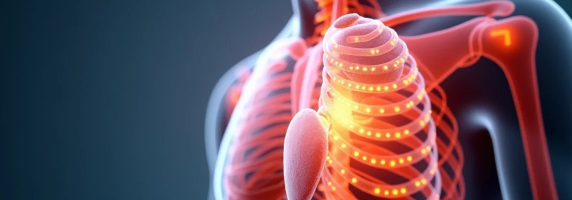
Anatomical structure and innervation of the right subcostal region
The anatomical complexity of the right subcostal region creates numerous opportunities for muscle spasm development. This area encompasses multiple tissue layers, from superficial intercostal muscles to deep visceral structures, each with distinct innervation patterns and functional relationships. Understanding these anatomical foundations proves essential for identifying the precise mechanisms underlying your muscle spasms and developing effective treatment strategies.
Intercostal muscle anatomy and fascia distribution
The intercostal muscles form three distinct layers between your ribs, with the external intercostals facilitating inspiration, the internal intercostals assisting expiration, and the innermost intercostals providing structural support. These muscles receive innervation from intercostal nerves, which originate from thoracic spinal cord segments T1-T12. When these nerves become irritated or compressed, they can trigger prolonged muscle contractions that manifest as spasms beneath your right rib cage.
The fascial connections in this region create a continuous network that can transmit tension and dysfunction across significant distances. The superficial fascia connects to deeper layers through fascial planes, potentially allowing localised muscle spasms to spread throughout the subcostal region. This fascial continuity explains why some patients experience widespread discomfort that seems disproportionate to the initial triggering event.
Phrenic nerve pathways and right hemidiaphragm connections
The right phrenic nerve, originating from cervical nerve roots C3-C5, provides motor innervation to the right hemidiaphragm and sensory innervation to the pericardium, pleura, and peritoneum. Dysfunction or irritation of this nerve can produce referred spasms in the subcostal region, particularly when diaphragmatic breathing patterns become compromised. The phrenic nerve’s extensive anatomical course makes it susceptible to various forms of mechanical and inflammatory irritation.
Diaphragmatic spasms often present as rhythmic contractions that may be mistaken for intercostal muscle spasms. These contractions can result from phrenic nerve hyperexcitability, electrolyte imbalances, or structural abnormalities affecting the nerve’s function. The close anatomical relationship between the diaphragm and subcostal muscles means that diaphragmatic dysfunction frequently triggers secondary muscle spasms in adjacent structures.
Hepatic capsule stretch receptors and visceral innervation
The liver’s fibrous capsule contains numerous stretch receptors that respond to changes in hepatic volume and pressure. When the liver becomes congested, inflamed, or enlarged, these receptors activate and can trigger reflex muscle spasms in the overlying intercostal and subcostal muscles. This viscerosomatic reflex represents a protective mechanism designed to limit movement and reduce mechanical stress on the compromised organ.
The hepatic innervation involves both sympathetic fibres from the coeliac plexus and parasympathetic fibres from the vagus nerve. Disruption of this complex innervation pattern can result in altered muscle tone and increased susceptibility to spasmodic episodes. The intricate relationship between visceral and somatic nervous systems explains why liver pathology frequently presents with significant muscular symptoms.
Thoracolumbar fascia attachments to lower ribs
The thoracolumbar fascia forms extensive attachments to the lower ribs, creating a mechanical link between the lumbar spine, pelvis, and thoracic cage. Dysfunction in the lumbar region can transmit tension through these fascial connections, potentially triggering muscle spasms in the subcostal area. This anatomical relationship explains why some patients with lower back problems develop secondary muscle spasms beneath their right ribs.
The multilayered structure of the thoracolumbar fascia provides attachment points for numerous muscles, including the latissimus dorsi, serratus posterior inferior, and internal obliques. When these muscles develop trigger points or become hypertonic, they can create sustained tension within the fascial system, predisposing you to recurrent spasmodic episodes in the subcostal region.
Musculoskeletal origins of right subcostal spasms
Musculoskeletal causes represent the most common source of muscle spasms beneath the right rib cage. These conditions typically involve direct muscle injury, overuse patterns, or biomechanical dysfunction that creates an environment conducive to involuntary muscle contractions. Understanding these mechanisms helps differentiate musculoskeletal causes from visceral pathology and guides appropriate treatment selection.
Intercostal muscle strain from respiratory overuse
Intercostal muscle strain commonly develops following intense physical activity, prolonged coughing episodes, or sudden respiratory movements. Athletes involved in sports requiring rapid breathing patterns or those with respiratory conditions may experience recurrent muscle spasms due to repetitive intercostal muscle overuse. The strain typically affects the external intercostal muscles most severely, as they bear the primary responsibility for inspiratory muscle work.
The development of muscle spasms following intercostal strain involves several physiological mechanisms. Initially, muscle fibre microtears trigger an inflammatory response that sensitises local nociceptors. This sensitisation can lead to protective muscle guarding, which may progress to sustained muscle contractions and eventual spasmodic activity. The body’s protective response often perpetuates the very problem it seeks to resolve , creating a cycle of muscle tension and spasm.
Serratus posterior inferior trigger point activation
The serratus posterior inferior muscle, which attaches to ribs 9-12, frequently develops trigger points that can refer pain and muscle spasms to the subcostal region. These trigger points typically form in response to poor posture, repetitive lifting activities, or sustained forward head positioning. When activated, these points can create both local muscle spasms and referred spasmodic activity in distant muscle groups.
Trigger point activation in the serratus posterior inferior creates a distinctive pattern of symptoms that includes deep, aching discomfort beneath the ribs and episodic muscle spasms that may worsen with breathing or movement. The referred pain pattern often extends from the trigger point location to the subcostal region, creating confusion about the true source of symptoms. Effective treatment requires addressing both the primary trigger points and any secondary muscle spasms they generate.
Quadratus lumborum referred pain patterns
The quadratus lumborum muscle, despite its primary location in the lumbar region, can generate referred pain and muscle spasms that extend to the subcostal area. This deep lumbar muscle plays a crucial role in lateral spinal flexion and hip hiking during gait. When trigger points develop within the quadratus lumborum, they can create complex referred pain patterns that include muscle spasms beneath the ribs.
The referred spasm pattern from quadratus lumborum dysfunction typically involves the lower intercostal muscles and may be accompanied by breathing difficulties during episodes. These spasms often occur in conjunction with movements that activate the quadratus lumborum, such as reaching overhead or lateral bending. The interconnected nature of the fascial system allows dysfunction in one region to manifest symptoms in seemingly unrelated areas.
Rib subluxation and costochondral junction dysfunction
Rib subluxations, particularly affecting the lower ribs, can trigger protective muscle spasms in the surrounding intercostal muscles. These subluxations may result from trauma, repetitive motion, or sudden twisting movements that exceed the normal range of motion at the costovertebral or costochondral joints. The resulting joint dysfunction creates mechanical irritation that the body attempts to protect through sustained muscle contractions.
Costochondral junction dysfunction represents a frequently overlooked cause of subcostal muscle spasms, particularly in individuals with hypermobile joints or those exposed to repetitive chest wall trauma.
When costochondral joints become dysfunctional, they can trigger both local and regional muscle spasms through mechanoreceptor activation and nociceptive input. The spasmodic response serves as a protective mechanism to limit further joint movement and prevent additional injury. However, this protective response can become self-perpetuating, leading to chronic muscle spasms that persist long after the initial joint dysfunction has resolved.
Hepatobiliary pathophysiology causing muscle spasms
The hepatobiliary system’s complex innervation patterns create numerous opportunities for visceral dysfunction to manifest as muscle spasms in the subcostal region. These organs share embryological origins and innervation pathways with the overlying musculature, creating viscerosomatic reflexes that can produce significant muscular symptoms. Understanding these relationships proves crucial for recognising when muscle spasms may indicate underlying organ dysfunction rather than primary musculoskeletal pathology.
Acute cholecystitis and gallbladder distension effects
Acute cholecystitis creates intense visceral nociceptive input that frequently triggers protective muscle spasms in the right subcostal region. The gallbladder’s rich innervation from sympathetic fibres originating at T7-T9 spinal levels creates direct neurological connections to the overlying intercostal muscles. When gallbladder inflammation develops, these shared neural pathways can produce referred muscle spasms that may precede or accompany the classic symptoms of cholecystitis.
Gallbladder distension, whether from acute cholecystitis or biliary obstruction, stretches mechanoreceptors within the organ wall and surrounding peritoneum. This mechanical stimulation activates viscerosomatic reflexes that increase muscle tone in segmentally related areas, including the right subcostal muscles. The resulting muscle spasms often exhibit a cramping quality and may intensify with deep inspiration or palpation of the affected area.
Hepatic congestion from right heart failure
Right heart failure leads to hepatic congestion, which can trigger muscle spasms through several mechanisms. The enlarged, congested liver stretches its fibrous capsule, activating stretch receptors that create reflex muscle guarding in the overlying subcostal region. Additionally, the increased pressure within the hepatic vessels can compress surrounding nerve structures, potentially triggering neuropathic muscle spasms.
The development of hepatic congestion often produces a characteristic pattern of muscle spasms that worsen with physical exertion or when lying flat. These spasms may be accompanied by other signs of right heart failure, such as peripheral oedema, jugular venous distension, and shortness of breath. The timing and character of these spasms can provide valuable diagnostic information about the underlying cardiac condition.
Biliary colic and smooth muscle contraction mechanisms
Biliary colic involves intense smooth muscle contractions within the biliary tree, typically triggered by gallstone impaction or biliary strictures. These contractions create visceral pain that can trigger secondary muscle spasms in the subcostal region through viscerosomatic reflexes. The smooth muscle contractions often occur in waves, producing episodic muscle spasms that mirror the underlying biliary dysfunction.
The neurological mechanisms underlying biliary colic involve both sympathetic and parasympathetic pathways, creating complex patterns of referred muscle activity. Sympathetic activation can increase muscle tone in segmentally related areas, while parasympathetic dysfunction may alter normal muscle coordination patterns. These neurological changes can persist beyond the acute episode, potentially contributing to chronic muscle spasm patterns in susceptible individuals.
Hepatomegaly-induced diaphragmatic irritation
Hepatomegaly can produce mechanical irritation of the right hemidiaphragm, triggering muscle spasms that extend throughout the subcostal region. The enlarged liver may directly contact the diaphragm or create tension within the supporting ligaments, both of which can activate mechanoreceptors and trigger reflex muscle contractions. This mechanical irritation often produces spasms that worsen with deep breathing or changes in position.
The relationship between hepatomegaly and diaphragmatic irritation demonstrates how organ enlargement can create widespread muscular symptoms through mechanical and neurological mechanisms.
Progressive hepatomegaly may also compress the inferior vena cava, creating venous congestion that can affect muscle metabolism and increase susceptibility to spasmodic episodes. The combination of mechanical irritation and metabolic dysfunction creates a complex pathophysiology that requires comprehensive treatment addressing both the underlying liver condition and the secondary muscular symptoms.
Gastrointestinal disorders triggering right rib spasms
Gastrointestinal pathology frequently manifests as muscle spasms in the subcostal region due to the extensive neural connections between the digestive system and the overlying musculature. The shared embryological development and innervation patterns of these structures create numerous opportunities for visceral dysfunction to produce somatic symptoms. These viscerosomatic relationships explain why gastrointestinal conditions often present with significant muscular components that may dominate the clinical picture.
Gastroesophageal reflux disease commonly triggers muscle spasms in the right subcostal area through several mechanisms. The presence of acid in the lower oesophagus activates nociceptors that can refer pain and muscle spasms to the chest wall and upper abdomen. Additionally, the chronic inflammation associated with GERD can sensitise neural pathways, making the musculature more susceptible to spasmodic episodes triggered by minor stimuli. The vagal innervation of the oesophagus creates direct connections to the overlying musculature, allowing oesophageal dysfunction to produce immediate muscular responses.
Small bowel obstruction or severe constipation can create significant abdominal distension that mechanically irritates the diaphragm and subcostal muscles. The increased intra-abdominal pressure alters normal respiratory mechanics, forcing the intercostal muscles to work harder and predisposing them to fatigue and spasmodic contractions. This mechanical relationship explains why gastrointestinal conditions often worsen during meals or when abdominal distension is most pronounced.
Inflammatory bowel conditions, such as Crohn’s disease or ulcerative colitis, can trigger muscle spasms through systemic inflammatory mediators that sensitise peripheral nociceptors. The chronic inflammatory state associated with these conditions can alter muscle membrane properties, making them more prone to spontaneous contractions. The systemic nature of inflammatory bowel disease often produces muscular symptoms that extend far beyond the primary site of gastrointestinal inflammation.
Peptic ulcer disease, particularly when involving the duodenum, can create referred muscle spasms in the right subcostal region through shared neural pathways. The deep visceral pain associated with duodenal ulceration often triggers protective muscle guarding that can evolve into sustained muscle spasms. The timing of these spasms, often correlating with meal patterns and gastric acid production, provides valuable diagnostic information about the underlying gastrointestinal pathology.
Respiratory system dysfunction and spasm development
Respiratory system dysfunction creates unique patterns of muscle spasms in the subcostal region due to the intimate relationship between breathing mechanics and intercostal muscle function. These muscles serve as primary or accessory muscles of respiration, making them particularly susceptible to dysfunction when respiratory patterns become compromised. Understanding these relationships proves essential for identifying respiratory causes of muscle spasms and developing appropriate treatment strategies that address both the muscular symptoms and underlying breathing dysfunction.
Chronic obstructive pulmonary disease significantly alters breathing patterns, forcing increased reliance on accessory respiratory muscles including the intercostals. The sustained increased workload on these muscles can lead to fatigue, trigger point formation, and eventual spasmodic episodes. The air trapping characteristic of COPD creates constant positive pressure within the thoracic cavity, altering the mechanical environment in which these muscles must function. This altered biomechanical environment predisposes the intercostal muscles to developing protective spasms, particularly during exacerbations of the underlying lung disease.
Asthma and other bronchospastic conditions can trigger muscle spasms through multiple mechanisms. The bronchospasm itself requires increased respiratory muscle work to overcome the increased airway resistance, potentially leading to muscle fatigue and spasmodic episodes. Additionally, the
widespread systemic inflammation associated with asthma can sensitise peripheral nociceptors throughout the thoracic region, making the intercostal muscles more prone to spasmodic contractions. The inflammatory mediators released during asthmatic episodes can directly affect muscle membrane properties, altering their electrical stability and increasing the likelihood of spontaneous contractions.
Pneumonia affecting the right lower lobe can create significant muscle spasms through both inflammatory and mechanical mechanisms. The infection triggers local inflammation that can irritate the overlying intercostal muscles and phrenic nerve branches. Additionally, the protective coughing associated with pneumonia places tremendous stress on the intercostal muscles, potentially leading to strain and subsequent spasmodic episodes. The combination of inflammation and mechanical stress creates a particularly challenging environment for muscle function in the subcostal region.
Pleuritic chest pain, whether from viral pleuritis or other causes, can trigger protective muscle spasms in the affected area. The sharp, stabbing pain characteristic of pleuritis often causes patients to alter their breathing patterns and adopt protective postures that can lead to secondary muscle dysfunction. The inflammatory process affecting the pleura can also directly irritate adjacent intercostal muscles through shared innervation pathways, creating a cycle of pain and muscle spasm that may persist beyond resolution of the primary pleural inflammation.
Sleep apnoea and other sleep-disordered breathing conditions can contribute to muscle spasms through chronic intermittent hypoxia and altered respiratory patterns. The frequent arousals and sleep fragmentation associated with sleep apnoea can disrupt normal muscle recovery processes, making the intercostal muscles more susceptible to spasmodic episodes. The chronic sympathetic activation associated with sleep apnoea can also alter muscle tone and increase baseline tension in the respiratory muscles, predisposing them to spasmodic contractions during periods of increased respiratory demand.
Differential diagnosis and clinical assessment protocols
Accurate diagnosis of muscle spasms beneath the right rib cage requires a systematic approach that distinguishes between musculoskeletal, visceral, and systemic causes. The overlapping symptom patterns and shared innervation pathways between different organ systems can create diagnostic challenges that require careful clinical assessment and targeted investigation. Developing an effective diagnostic strategy involves understanding the characteristic features of each potential cause and implementing appropriate testing protocols to confirm or exclude specific conditions.
The initial clinical assessment should focus on obtaining a detailed history that explores the onset, character, and aggravating factors of the muscle spasms. Sudden onset spasms following physical activity or trauma typically suggest musculoskeletal causes, while gradual onset spasms associated with systemic symptoms may indicate visceral pathology. The timing of symptoms relative to meals, breathing patterns, and body position provides valuable diagnostic information that can help differentiate between various potential causes.
A thorough understanding of the temporal patterns and associated symptoms is crucial for distinguishing between musculoskeletal and visceral causes of right subcostal muscle spasms.
Physical examination should include assessment of the chest wall, abdomen, and spine to identify areas of muscle tension, trigger points, or structural abnormalities. The hooking maneuver test can help identify slipping rib syndrome, while palpation of the subcostal region during different breathing phases can reveal patterns consistent with diaphragmatic dysfunction. Neurological testing should assess intercostal nerve function and identify any signs of nerve compression or irritation that might contribute to muscle spasm development.
Laboratory investigations should be tailored to the suspected underlying cause, with liver function tests and inflammatory markers helping to identify hepatobiliary pathology or systemic inflammatory conditions. Cardiac enzymes and electrocardiography may be necessary when cardiac causes are suspected, particularly in patients with risk factors for heart disease. The selection of appropriate diagnostic tests depends heavily on the clinical presentation and identified risk factors during the initial assessment.
Imaging studies play a crucial role in the diagnostic evaluation, with chest X-rays helping to identify pulmonary pathology or rib abnormalities. Abdominal ultrasound can assess for hepatobiliary pathology, while CT scanning may be necessary to evaluate complex cases or when multiple organ systems are potentially involved. MRI of the thoracic spine may be indicated when nerve compression or structural abnormalities are suspected as contributing factors to the muscle spasm development.
Electromyographic studies can provide valuable information about muscle function and help identify specific patterns of muscle activation that may contribute to spasmodic episodes. These studies can also help differentiate between primary muscle disorders and secondary muscle dysfunction resulting from nerve dysfunction or central nervous system pathology. The timing and coordination of muscle contractions revealed by EMG studies can guide targeted treatment approaches and monitor treatment response over time.
Response to therapeutic interventions can provide important diagnostic information, with muscle spasms responding to anti-inflammatory medications potentially indicating inflammatory causes, while spasms responding to muscle relaxants suggesting primary muscle pathology. The response to positional changes, breathing exercises, or specific manual therapy techniques can help identify mechanical causes and guide ongoing treatment strategies. Understanding how different interventions affect symptom patterns provides valuable insight into the underlying pathophysiology and helps refine the diagnostic assessment.
The differential diagnosis must also consider less common but important causes such as intercostal neuralgia, thoracic outlet syndrome, or referred pain from cervical spine pathology. These conditions can produce similar symptom patterns but require different treatment approaches, making accurate differentiation crucial for optimal patient outcomes. The complexity of the anatomical relationships in the thoracic region means that multiple contributing factors may be present simultaneously, requiring a comprehensive assessment approach that addresses all potential causes rather than focusing on a single suspected pathology.