Discovering a lump under the breast near the ribs can be an unsettling experience that triggers immediate concern. This anatomical region, known as the lateral chest wall or inframammary area, represents a complex intersection of breast tissue, chest wall structures, and various anatomical components. Understanding the potential causes of lumps in this specific location requires knowledge of the intricate anatomy and the diverse pathological processes that can affect this region.
The area beneath the breast and adjacent to the ribcage encompasses multiple tissue types, including mammary tissue extensions, muscular structures, fascial planes, and subcutaneous layers. Each of these components can give rise to various types of masses, ranging from benign conditions to more serious pathological processes. Early recognition and proper evaluation of any new lumps in this region remain crucial for appropriate medical management and peace of mind.
While many lumps discovered in this location prove to be benign, the complexity of the anatomical region necessitates thorough understanding of potential causes. From simple lipomas to complex inflammatory processes, the differential diagnosis for lumps under the breast near the ribs spans a broad spectrum of conditions that require careful consideration and often professional medical evaluation.
Anatomical structure of the chest wall and intercostal region
The chest wall represents a sophisticated architectural framework comprising multiple layers of tissues, each with distinct anatomical characteristics and potential pathological processes. Understanding this complex anatomy proves essential for comprehending how various types of lumps can develop in the region beneath the breast and adjacent to the ribs. The chest wall consists of three primary layers: the superficial layer containing skin and subcutaneous fat, the intermediate layer housing the shoulder girdle and pectoralis muscles, and the deep layer encompassing the bony chest structures including sternum, ribs, and thoracic spine.
The intercostal spaces between adjacent ribs contain neurovascular bundles, intercostal muscles, and fascial planes that can become sites of pathological processes. These spaces provide pathways for nerve transmission, blood circulation, and lymphatic drainage, making them susceptible to various inflammatory and neoplastic conditions. The intricate relationship between these anatomical components creates multiple potential sites where lumps can develop, each with characteristic presentations and implications.
Pectoralis major and minor muscle anatomy
The pectoralis major muscle forms the primary muscular component of the anterior chest wall, originating from the clavicle, sternum, and upper costal cartilages before inserting into the humerus. This large, fan-shaped muscle can develop various pathological conditions including strain injuries, haematomas, and rarely, primary muscle tumours. Beneath the pectoralis major lies the smaller pectoralis minor muscle, which extends from the third to fifth ribs to the coracoid process of the scapula.
Muscular injuries or inflammatory processes affecting these structures can present as palpable masses in the lateral chest region. Post-exercise muscle swelling, traumatic haematomas, or inflammatory myositis can create temporary or persistent lumps that may be mistaken for more serious pathological processes. Understanding the anatomical boundaries and functional characteristics of these muscles helps differentiate muscular causes from other potential sources of chest wall masses.
Intercostal muscle groups and fascial layers
The intercostal spaces contain three distinct muscle layers: the external intercostals, internal intercostals, and innermost intercostals. These muscles play crucial roles in respiratory mechanics and chest wall stability. Between these muscle layers run the intercostal vessels and nerves, creating neurovascular bundles that follow predictable anatomical pathways. Pathological processes affecting these structures can manifest as localised swelling or mass formation.
Fascial layers surrounding these muscles create potential spaces where fluid collections, haematomas, or inflammatory processes can accumulate. The endothoracic fascia, which lines the inner surface of the chest wall, represents an important anatomical landmark that separates chest wall pathology from pleural or pulmonary processes. Understanding these fascial relationships proves crucial for accurate diagnosis and appropriate treatment planning when evaluating chest wall lumps.
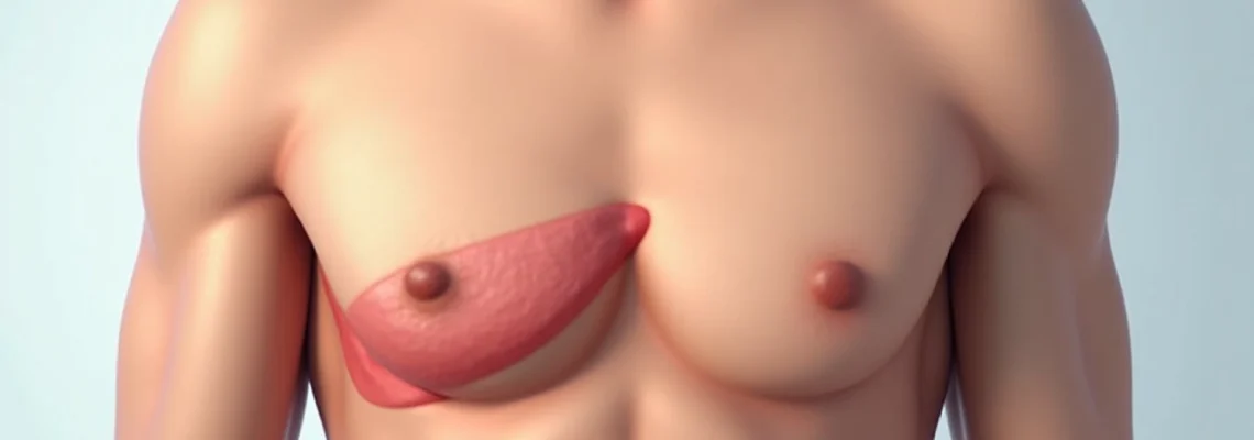
Mammary ridge development and supernumerary breast tissue
During embryological development, the mammary ridge extends from the axilla to the inguinal region, with most portions regressing except for the area destined to become the normal breast. Occasionally, remnants of this embryological structure persist, creating supernumerary breast tissue or accessory nipples along the mammary line. These developmental variants most commonly occur in the axillary region but can extend to areas beneath the normal breast.
Supernumerary breast tissue can undergo the same pathological processes as normal mammary tissue, including hormonal changes, inflammatory conditions, and neoplastic transformation. During pregnancy, lactation, or hormonal fluctuations, these tissue remnants may become more prominent and palpable, potentially causing confusion about their nature and significance. Recognition of these developmental variants prevents unnecessary anxiety and ensures appropriate management when they become clinically apparent.
Costal cartilage and sternocostal joint positioning
The costal cartilages connect the ribs to the sternum, forming the sternocostal joints that allow for chest wall movement during respiration. These cartilaginous structures can become sites of inflammatory processes such as costochondritis or Tietze syndrome, leading to localised swelling and tenderness. The anatomical position of these joints, particularly those of the lower ribs, places them in close proximity to the inframammary region.
Pathological processes affecting the costal cartilages can create palpable masses that may be mistaken for breast or soft tissue lumps. Understanding the normal anatomical landmarks and the potential for cartilaginous inflammation helps differentiate these musculoskeletal causes from other pathological processes. The relationship between chest wall movement and symptom reproduction often provides important diagnostic clues when evaluating lumps in this region.
Benign breast masses in the lateral chest region
Benign breast masses represent the most common cause of lumps discovered in the lateral chest region beneath the breast. These non-cancerous growths encompass a diverse range of histological types, each with characteristic features, risk factors, and clinical presentations. Understanding the various types of benign breast masses helps provide reassurance while ensuring appropriate evaluation and management strategies.
The lateral extent of breast tissue often extends beyond what might be considered the traditional breast boundary, particularly in the axillary tail region and inferior breast quadrants. This anatomical variation means that typical breast pathology can present in areas that might initially appear to be outside the breast proper. Hormonal influences, genetic factors, and environmental exposures all contribute to the development of various benign breast conditions that can manifest as palpable lumps in the lateral chest region.
Fibroadenoma characteristics and hormonal influences
Fibroadenomas represent the most common benign breast tumours, particularly affecting women in their reproductive years. These well-circumscribed masses consist of both glandular and stromal components, creating firm, mobile lumps that can occur anywhere within breast tissue, including the lateral regions. Fibroadenomas typically feel smooth and rubbery, with clearly defined borders that allow them to move freely within the surrounding breast tissue.
Hormonal fluctuations significantly influence fibroadenoma behaviour, with many masses showing cyclical changes in size and tenderness corresponding to menstrual cycles. During pregnancy and lactation, fibroadenomas may undergo rapid growth due to increased hormonal stimulation. The vast majority of fibroadenomas remain stable over time , though some may gradually decrease in size, particularly after menopause when hormonal influences diminish.
Lipoma formation in subcutaneous tissue
Lipomas represent benign tumours composed of mature adipose tissue that can develop anywhere in the body where fat cells exist. In the chest wall region, lipomas commonly occur in the subcutaneous layer, creating soft, mobile masses that may be located beneath or adjacent to breast tissue. These growths typically develop slowly over time and rarely cause symptoms unless they reach considerable size or compress adjacent structures.
The characteristic features of lipomas include their soft, doughy consistency and excellent mobility within the surrounding tissues. Most lipomas remain stable in size over extended periods, though some may gradually enlarge. The benign nature of most lipomas means that treatment is typically only necessary when they cause cosmetic concerns, discomfort, or uncertainty about diagnosis. Surgical excision provides definitive treatment when removal becomes necessary.
Sebaceous cyst development and keratin accumulation
Sebaceous cysts, more accurately termed epidermoid cysts, develop when sebaceous glands become blocked, leading to accumulation of keratin and sebaceous material within a cystic cavity. These common benign lesions can occur anywhere on the chest wall where sebaceous glands exist, including areas beneath and adjacent to the breast. The characteristic features include a central punctum or opening and a distinctive odour when the cyst contents are expressed.
Most sebaceous cysts remain asymptomatic unless they become infected or inflamed, at which point they may become tender, red, and enlarged. The cyst wall typically prevents complete drainage without surgical intervention, meaning that simple incision and drainage often results in recurrence. Complete surgical excision including the cyst wall provides definitive treatment and prevents recurrence, though this is typically only necessary when cysts become symptomatic or infected.
Galactocele formation during lactation period
Galactoceles represent milk-filled cysts that develop during or shortly after the lactation period when milk ducts become obstructed. These benign lesions can occur anywhere within breast tissue, including the lateral and inferior regions that extend beneath the traditional breast boundaries. Galactoceles typically present as smooth, mobile masses that may fluctuate in size depending on breastfeeding patterns and milk production.
The characteristic history of recent pregnancy or lactation, combined with the typical physical findings, usually makes diagnosis straightforward. Most galactoceles resolve spontaneously as lactation ceases and hormonal influences diminish. Persistent or symptomatic galactoceles may require aspiration or surgical intervention, though conservative management is often successful. Understanding the relationship between lactation and galactocele formation helps provide appropriate reassurance and management strategies.
Malignant neoplasms and breast cancer manifestations
While benign conditions represent the majority of lumps discovered beneath the breast near the ribs, malignant neoplasms constitute an important differential consideration that requires careful evaluation. Breast cancer can present in various forms and locations, including the lateral and inferior aspects of the breast where tissue extends toward the chest wall. Understanding the characteristics and presentation patterns of malignant breast disease proves crucial for ensuring appropriate evaluation and timely intervention when necessary.
The anatomical extent of breast tissue means that malignant processes can manifest in areas that might initially appear to be outside the traditional breast boundaries. Additionally, the rich lymphatic drainage of the breast region means that metastatic disease from other primary sites can occasionally present as masses in the lateral chest wall. Early detection and accurate diagnosis remain paramount for optimal outcomes when dealing with malignant processes in this region.
Invasive ductal carcinoma presentation patterns
Invasive ductal carcinoma represents the most common form of breast cancer, accounting for approximately 80% of all breast malignancies. These tumours can develop anywhere within breast tissue, including the lateral and inferior quadrants that extend toward the chest wall. Unlike benign masses, invasive ductal carcinomas typically feel firm to hard, with irregular borders and limited mobility due to their tendency to infiltrate surrounding tissues.
The characteristic features that may suggest malignancy include fixation to underlying structures, skin changes such as dimpling or peau d’orange appearance, and associated lymphadenopathy. The absence of pain does not exclude malignancy , as many breast cancers present as painless masses. Associated symptoms such as nipple discharge, skin retraction, or changes in breast contour warrant immediate medical evaluation regardless of the location of any palpable abnormalities.
Inflammatory breast cancer clinical features
Inflammatory breast cancer represents an aggressive form of breast malignancy that can present with diffuse inflammation and swelling rather than a discrete palpable mass. This rare but serious condition can affect any area of the breast, including the lateral and inferior regions adjacent to the chest wall. The characteristic presentation includes rapid onset of breast swelling, redness, warmth, and skin changes resembling an orange peel texture.
Unlike typical breast cancers that present as discrete lumps, inflammatory breast cancer often manifests as diffuse breast enlargement and skin changes that may initially be mistaken for infectious processes. The rapid progression of symptoms, typically occurring over weeks rather than months or years, helps distinguish this condition from benign inflammatory processes. The aggressive nature of inflammatory breast cancer necessitates immediate medical evaluation and prompt initiation of treatment when this diagnosis is suspected.
Lymphoma development in breast tissue
Primary breast lymphomas represent rare malignancies that can develop within breast tissue or adjacent lymph nodes. These tumours may present as rapidly growing masses anywhere within the breast, including the lateral regions near the chest wall. The presentation often includes painless, firm masses that may be associated with systemic symptoms such as fever, night sweats, and unintentional weight loss.
Secondary involvement of the breast by systemic lymphomas is more common than primary breast lymphomas, particularly in patients with known lymphomatous disease. The characteristic features may include multiple masses, rapid growth, and association with enlarged lymph nodes in other regions. The distinction between primary and secondary breast lymphomas requires comprehensive evaluation including appropriate imaging studies and tissue sampling for definitive diagnosis.
Metastatic disease from primary sites
Metastatic disease to the breast from other primary sites represents an uncommon but important consideration when evaluating breast masses. The most frequent primary sites include lung, melanoma, gastrointestinal tract, and gynaecological organs. Metastatic deposits can occur anywhere within breast tissue, including the lateral and inferior regions adjacent to the chest wall.
The clinical presentation of metastatic disease to the breast varies depending on the primary tumour type and the extent of systemic disease. Some metastatic deposits present as well-circumscribed masses that may be difficult to distinguish from primary breast pathology, while others may cause more diffuse changes. A history of previous malignancy significantly increases the suspicion for metastatic disease, though metastases can occasionally represent the initial presentation of an occult primary tumour.
Musculoskeletal conditions affecting the ribcage area
The complex musculoskeletal anatomy of the chest wall creates multiple potential sites where various pathological processes can manifest as palpable lumps or masses. These conditions often arise from the bony structures of the ribcage, the intercostal muscles, or the various fascial planes that comprise the chest wall architecture. Understanding these musculoskeletal causes proves essential for accurate diagnosis and appropriate management, particularly since many of these conditions can mimic more serious pathological processes.
Musculoskeletal conditions affecting the ribcage area often present with characteristic features that help distinguish them from breast-related pathology. These may include relationships to physical activity, respiratory movements, or specific positional changes. The proximity of chest wall structures to breast tissue means that musculoskeletal pathology can often be mistaken for breast-related conditions, making careful evaluation and understanding of anatomical relationships crucial for accurate diagnosis.
Costochondritis represents one of the most common musculoskeletal causes of chest wall discomfort and apparent mass formation. This inflammatory condition affects the cartilaginous connections between the ribs and sternum, leading to localised swelling, tenderness, and pain that may worsen with movement or deep inspiration. The inflammatory process can create palpable thickening along the affected costal cartilages, particularly those of the middle and lower ribs that lie in close proximity to the inframammary region.
Tietze syndrome represents a related but distinct condition characterised by painful swelling of one or more costal cartilages, most commonly affecting the second and third ribs. Unlike costochondritis, Tietze syndrome typically presents with visible and palpable swelling that persists for extended periods. The affected cartilages may remain enlarged for months or even years, creating persistent palpable masses along the chest wall. The inflammatory nature of these conditions means that symptoms often fluctuate in intensity and may be exacerbated by physical activity, respiratory infections, or stress.
Rib fractures, whether traumatic or pathological, can result in callus formation and bony irregularities that present as palpable lumps along the chest wall. Healing rib fractures typically develop fibrous and eventually bony callus tissue that serves to stabilise the fracture site. This healing process can create
permanent palpable lumps that may persist long after the initial injury has healed. In cases of multiple rib fractures or complex chest wall trauma, the healing process can result in significant chest wall deformity and multiple palpable irregularities that require careful evaluation to distinguish from pathological masses.
Intercostal muscle strains or tears can result in haematoma formation within the muscle belly or at the musculotendinous junction. These injuries commonly occur following vigorous physical activity, persistent coughing, or direct trauma to the chest wall. The resulting haematomas can present as tender, palpable masses within the intercostal spaces that may initially cause concern about more serious pathological processes. The relationship between symptom onset and physical activity often provides important diagnostic clues, particularly when combined with characteristic pain patterns that worsen with specific movements or respiratory efforts.
Fibrous dysplasia represents a benign bone disorder that can affect the ribs, leading to localised bone expansion and potential mass formation. This condition involves replacement of normal bone with fibrous tissue, creating areas of weakness and potential deformity. While most cases remain asymptomatic, larger lesions can present as palpable lumps along the affected ribs. The gradual, painless enlargement over time helps distinguish fibrous dysplasia from acute inflammatory processes or traumatic injuries.
Infectious and inflammatory processes
Infectious and inflammatory processes affecting the chest wall can manifest as various types of lumps and masses, ranging from superficial skin infections to deep-seated inflammatory conditions involving the chest wall structures. These processes can arise from direct inoculation through skin breaks, haematogenous spread from distant sites, or extension from adjacent infectious foci. Understanding the spectrum of infectious causes proves crucial for appropriate recognition and treatment, particularly since delayed diagnosis can lead to serious complications.
The rich vascular supply and lymphatic drainage of the chest wall region makes it susceptible to various infectious processes that can present as rapidly developing masses. The clinical presentation often includes systemic signs of infection such as fever, malaise, and elevated inflammatory markers, helping to distinguish infectious causes from benign developmental or neoplastic processes. The temporal relationship between symptom onset and potential predisposing factors provides important diagnostic information.
Cellulitis represents a common superficial soft tissue infection that can affect the skin and subcutaneous tissues of the chest wall. This bacterial infection typically presents with erythema, warmth, swelling, and tenderness that may initially appear as a localised mass or lump. The infection commonly follows minor skin trauma, insect bites, or breaks in skin integrity that allow bacterial entry. Streptococcal and staphylococcal organisms represent the most frequent causative agents, though other bacteria may be involved in immunocompromised patients or following specific exposure risks.
Chest wall abscesses can develop as complications of cellulitis or arise from haematogenous seeding of susceptible tissues. These collections of purulent material present as fluctuant, tender masses that may be associated with systemic signs of infection. The characteristic features include central fluctuance surrounded by inflammatory changes and potential skin breakdown with spontaneous drainage. Successful treatment typically requires both antimicrobial therapy and surgical drainage, particularly for larger collections or those failing to respond to conservative management.
Hidradenitis suppurativa represents a chronic inflammatory condition affecting apocrine sweat glands that can involve the chest wall region, particularly in areas with hair follicles and sweat glands. This condition presents with recurrent painful nodules, abscesses, and eventually sinus tract formation in affected areas. The chronic nature of the condition, with periods of exacerbation and remission, helps distinguish it from acute infectious processes. Treatment often requires long-term management strategies including antimicrobial therapy, anti-inflammatory medications, and sometimes surgical intervention for severe cases.
Osteomyelitis of the ribs or sternum represents a serious infectious condition that can present with localised bone pain and potential mass formation due to periosteal reaction and soft tissue inflammation. This condition may arise from haematogenous spread, direct inoculation following trauma or surgery, or extension from adjacent soft tissue infections. The deep-seated nature of bone infections often results in persistent, aching pain that worsens progressively over time. Imaging studies typically reveal characteristic bone changes including cortical destruction, periosteal reaction, and associated soft tissue inflammation that help confirm the diagnosis.
Tuberculous involvement of the chest wall, while uncommon in developed countries, remains an important consideration in certain populations and geographic regions. Tuberculous chest wall infections can present as chronic, slowly progressive masses that may be associated with sinus tract formation and characteristic caseous drainage. The indolent course and potential for multi-organ involvement distinguish tuberculous infections from more acute bacterial processes. Diagnosis requires specific microbiological testing and often tissue biopsy for acid-fast bacilli identification.
Diagnostic imaging protocols and differential assessment
Comprehensive evaluation of lumps beneath the breast near the ribs requires systematic diagnostic approaches that combine clinical assessment with appropriate imaging modalities. The selection of imaging techniques depends on multiple factors including the clinical presentation, patient age, size and characteristics of the palpable abnormality, and the suspected differential diagnosis. Modern imaging protocols provide detailed anatomical information that helps distinguish between various pathological processes and guides appropriate management decisions.
Initial clinical evaluation remains fundamental to the diagnostic process, involving detailed history taking and thorough physical examination. The temporal evolution of the lump, associated symptoms, relationship to menstrual cycles, family history, and previous medical conditions all provide crucial diagnostic information. Physical examination techniques including inspection, palpation, and assessment of mobility help characterise the mass and identify associated findings such as skin changes, lymphadenopathy, or chest wall abnormalities.
Mammography represents the standard screening and diagnostic imaging modality for breast-related masses in appropriate age groups. Digital mammography with tomosynthesis provides detailed visualisation of breast tissue architecture and can identify masses, architectural distortion, and microcalcifications that may not be apparent on physical examination. However, mammographic evaluation of the extreme lateral and inferior breast regions can be technically challenging, sometimes requiring specialised projections or supplementary imaging modalities for complete assessment.
Breast ultrasonography serves as an essential complement to mammographic evaluation, particularly for characterising palpable abnormalities and distinguishing solid masses from cystic lesions. The real-time nature of ultrasound examination allows correlation with palpable findings and provides detailed information about mass characteristics including echogenicity, vascularity, and relationship to surrounding structures. Doppler ultrasound assessment of blood flow patterns can provide additional information about the nature of identified masses, with increased vascularity often suggesting active pathological processes.
Magnetic resonance imaging of the breast offers superior soft tissue contrast and can provide detailed information about the extent of pathological processes, particularly in cases where mammography and ultrasound findings are inconclusive. MRI proves particularly valuable for evaluating complex masses, assessing for multifocal or multicentric disease, and characterising enhancement patterns that may suggest malignancy. The ability to evaluate both breasts simultaneously and assess chest wall involvement makes MRI valuable for comprehensive evaluation of lateral chest wall masses.
Computed tomography scanning provides excellent visualisation of chest wall bony structures and can identify pathological processes involving the ribs, intercostal spaces, and deeper chest wall structures. CT imaging proves particularly valuable for evaluating suspected musculoskeletal causes of chest wall masses and can provide detailed information about bone integrity, soft tissue involvement, and relationship to adjacent structures. Contrast-enhanced CT studies can help differentiate infectious processes from neoplastic conditions based on enhancement patterns and associated findings.
When imaging studies suggest concerning features or when clinical suspicion remains high despite reassuring imaging findings, tissue sampling through various biopsy techniques provides definitive diagnostic information. Core needle biopsy under image guidance offers a minimally invasive method for obtaining adequate tissue samples for histopathological evaluation. The choice of guidance modality depends on lesion characteristics and visibility with different imaging techniques, with ultrasound guidance being preferred for most palpable masses due to its real-time capabilities and lack of ionising radiation.
Fine needle aspiration cytology may be appropriate for certain cystic lesions or when core biopsy is technically challenging, though the limited tissue architecture information obtained through cytology may require subsequent core biopsy for definitive diagnosis. The correlation between imaging findings, clinical presentation, and histopathological results ensures appropriate diagnostic accuracy and helps guide subsequent management decisions. Multidisciplinary consultation involving radiologists, clinicians, and pathologists optimises diagnostic accuracy and treatment planning for complex cases.
Follow-up protocols vary depending on the specific diagnosis and clinical circumstances, with benign conditions often requiring periodic monitoring to ensure stability while more concerning findings necessitate prompt treatment initiation. The development of standardised imaging and reporting systems helps ensure consistent evaluation and communication between healthcare providers, ultimately improving patient outcomes through systematic diagnostic approaches and appropriate management strategies.