Triple arthrodesis represents one of the most significant reconstructive procedures in foot and ankle surgery, fundamentally altering the biomechanics of the hindfoot through fusion of three critical joints: the subtalar, talonavicular, and calcaneocuboid joints. This comprehensive surgical intervention addresses severe deformities, arthritis, and instability that cannot be managed through conservative treatment or less invasive procedures. While the surgery effectively eliminates pain and corrects deformity, patients embarking on this journey must understand the profound changes that will occur in their foot function, gait patterns, and daily activities.
The decision to undergo triple arthrodesis is never taken lightly, as it involves permanently sacrificing the natural motion of the hindfoot complex in exchange for stability and pain relief. Modern surgical techniques have significantly improved outcomes, yet the recovery process remains lengthy and demanding. Understanding what life looks like after this procedure helps patients set realistic expectations and prepare for the rehabilitation journey ahead.
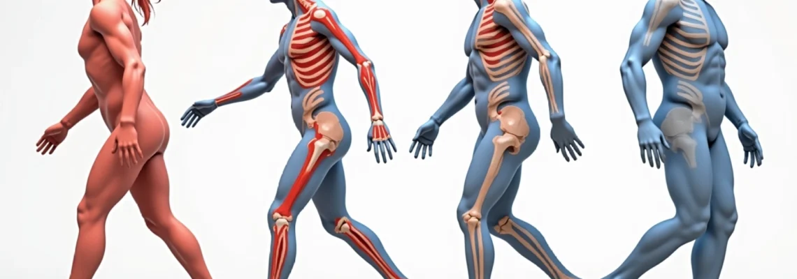
Understanding triple arthrodesis: talonavicular, subtalar, and calcaneocuboid joint fusion
Triple arthrodesis fundamentally transforms the architecture of the foot by eliminating motion in the three joints that collectively comprise the hindfoot complex. The talonavicular joint, positioned at the apex of the medial longitudinal arch, plays a crucial role in foot pronation and supination. When fused, this joint loses its ability to accommodate ground irregularities and adapt to changing terrain. The subtalar joint, responsible for inversion and eversion movements, becomes immobilised, dramatically reducing the foot’s capacity for side-to-side adjustments during walking.
The calcaneocuboid joint, forming part of the lateral column, contributes to the foot’s length and stability during weight-bearing activities. Its fusion creates a rigid lateral border that cannot flex or adapt during the gait cycle. Together, these three fusions create what surgeons call a tripod effect , where the foot becomes a stable but inflexible platform for weight bearing.
Pantalar joint complex biomechanical changes Post-Fusion
Following triple arthrodesis, the pantalar joint complex undergoes significant biomechanical restructuring. The ankle joint, or tibiotalar articulation, must now compensate for the loss of hindfoot motion by increasing its range of movement in both sagittal and coronal planes. This compensation mechanism places additional stress on the ankle joint surfaces, potentially accelerating degenerative changes over time. Research indicates that up to 77% of patients develop some degree of ankle arthritis within 15 years following triple arthrodesis.
The elimination of subtalar motion forces the ankle to accommodate rotational forces that were previously shared between multiple joints. This altered load distribution can lead to increased pressure on specific areas of the tibiotalar joint, particularly during activities requiring directional changes or movement on uneven surfaces. The ankle joint’s cartilage, not designed to handle these additional stresses long-term, may begin showing signs of wear within the first few years post-surgery.
Hindfoot and midfoot motion compensation mechanisms
The human foot demonstrates remarkable adaptability when faced with the loss of hindfoot motion. Following triple arthrodesis, the midfoot joints, particularly the naviculocuneiform and tarsometatarsal articulations, increase their range of motion to partially compensate for the fused hindfoot. This compensation comes at a cost, as these joints experience significantly higher loads than their natural design intended. Studies show that midfoot arthritis develops in approximately 56% of patients within seven years of triple arthrodesis.
The transverse tarsal joint, comprising the talonavicular and calcaneocuboid joints, normally works in concert with the subtalar joint to provide foot flexibility. With two-thirds of this complex now fused, the remaining mobile segments must work harder to maintain functional foot mechanics. This increased demand on the unfused portions of the transverse tarsal joint contributes to accelerated wear and potential future surgical intervention.
Impact on tibiotalar joint function and ankle dorsiflexion
Ankle dorsiflexion, the upward movement of the foot towards the shin, becomes particularly important following triple arthrodesis. With hindfoot motion eliminated, the ankle joint must provide all sagittal plane movement required for activities such as walking uphill, climbing stairs, and squatting. Many patients notice that achieving adequate dorsiflexion becomes more challenging, not due to ankle stiffness, but because the foot can no longer assist through hindfoot motion.
The tibiotalar joint experiences altered contact pressures post-fusion, with studies using dynamic foot models showing increased peak loading pressure, particularly in the anterior aspect of the ankle. This pressure redistribution can lead to localised cartilage damage and the development of osteophytes or bone spurs. Patients may experience anterior ankle impingement symptoms, characterised by pain during dorsiflexion activities.
Adjacent joint disease development in chopart and lisfranc joints
The Chopart joint complex, encompassing the talonavicular and calcaneocuboid articulations, bears the brunt of compensatory changes following triple arthrodesis. With the talonavicular joint fused and the calcaneocuboid joint immobilised, the remaining mobile structures within the midfoot must accommodate rotational and adaptive movements. The naviculocuneiform joints, in particular, experience increased loading that can lead to degenerative changes within five to ten years post-surgery.
Lisfranc joint arthritis develops as a consequence of the rigid hindfoot creating altered ground reaction forces during weight bearing. The tarsometatarsal joints, designed to provide fine-tuning adjustments during gait, become overloaded when the hindfoot cannot contribute to shock absorption and terrain adaptation. This phenomenon explains why approximately 44% of patients develop Lisfranc joint arthritis following triple arthrodesis, often requiring additional surgical intervention years later.
Immediate Post-Operative recovery timeline and surgical wound management
The immediate post-operative period following triple arthrodesis requires meticulous attention to wound healing and bone fusion processes. The extensive soft tissue dissection necessary for this procedure creates significant swelling and potential complications that must be carefully managed. Patients typically remain hospitalised for two to four days, during which elevation of the operative extremity above heart level remains crucial for minimising oedema and promoting optimal healing conditions.
Wound management involves monitoring multiple incision sites, as triple arthrodesis typically requires both medial and lateral surgical approaches. The lateral incision, in particular, requires careful observation due to the relatively poor blood supply in this area. Surgeons may employ negative pressure wound therapy or specialised dressing techniques to optimise healing in patients with compromised circulation or diabetes.
Non-weight bearing phase: weeks 1-8 cast immobilisation protocol
The initial non-weight bearing phase represents the most critical period for successful bone fusion. Patients must remain completely non-weight bearing on the operative extremity for a minimum of six weeks, with many surgeons extending this period to eight weeks depending on individual healing factors. This extended immobilisation period allows the surgically prepared bone surfaces to begin the complex process of osseous union without mechanical disruption.
Cast immobilisation during this phase serves multiple purposes beyond simple bone protection. The rigid immobilisation prevents micro-motion at the fusion sites, which could disrupt the delicate early callus formation. Additionally, the cast provides compression that helps control post-operative swelling and maintains optimal alignment of the fused joints. Patients typically require cast changes at two-week intervals to accommodate decreasing swelling and ensure proper fit.
Compliance with non-weight bearing restrictions proves challenging for many patients, particularly those accustomed to active lifestyles. The use of assistive devices such as knee scooters , hands-free crutches, or wheelchair adaptation becomes essential for maintaining independence during this period. Physical therapy during the non-weight bearing phase focuses on maintaining strength in the unaffected extremities and preventing complications such as blood clots or muscle atrophy.
Radiographic bone healing assessment using CT and plain radiographs
Monitoring bone healing progression requires a systematic approach combining clinical assessment with advanced imaging techniques. Plain radiographs remain the initial screening tool, providing essential information about hardware positioning, overall alignment, and early signs of bone union. However, the complex three-dimensional nature of triple arthrodesis often necessitates computed tomography scanning for definitive assessment of fusion status.
CT scanning proves particularly valuable in detecting non-union or delayed union, which occurs in approximately 5-11% of triple arthrodesis procedures. The cross-sectional imaging allows surgeons to evaluate each fusion site independently, identifying areas of incomplete bone healing that may not be apparent on standard radiographs. This detailed assessment guides decisions about weight-bearing progression and the potential need for additional interventions.
Pin site care and external fixation device maintenance
Some triple arthrodesis procedures utilise external fixation devices or prominent percutaneous pins for additional stability during the healing phase. These hardware elements require specific care protocols to prevent infection and maintain optimal function. Pin site care involves daily cleaning with saline solution or prescribed antiseptic solutions, along with monitoring for signs of inflammation, drainage, or loosening.
External fixation frames, while less commonly used in routine triple arthrodesis, may be employed in complex deformity corrections or revision procedures. Patients with external fixators require education about device care, including proper cleaning techniques, recognition of potential complications, and activity modifications necessary to prevent hardware failure. The psychological impact of visible external hardware should not be underestimated, as many patients experience anxiety about the appearance and function of these devices.
Thromboembolism prevention with rivaroxaban and compression therapy
The combination of surgical trauma, prolonged immobilisation, and non-weight bearing status significantly increases the risk of venous thromboembolism following triple arthrodesis. Modern prophylaxis protocols often incorporate direct oral anticoagulants such as rivaroxaban, which provide effective clot prevention without the need for frequent monitoring required with traditional warfarin therapy. The typical prophylaxis course extends for 10-14 days post-operatively, though high-risk patients may require extended treatment.
Mechanical prophylaxis through compression therapy plays an equally important role in thromboembolism prevention. Graduated compression stockings applied to the non-operative extremity help maintain venous return, while pneumatic compression devices may be utilised during the immediate post-operative period. Patient education about recognising symptoms of deep vein thrombosis or pulmonary embolism remains crucial, as early detection and treatment can prevent life-threatening complications.
Long-term functional outcomes and gait pattern adaptations
The long-term functional outcomes following triple arthrodesis demonstrate significant improvements in pain relief and patient satisfaction, despite measurable changes in foot and ankle biomechanics. Studies with 15-year follow-up periods indicate that 87-95% of patients report satisfaction with their surgical outcome and would choose to undergo the procedure again. However, these positive outcomes come with notable functional adaptations that patients must understand and accommodate in their daily lives.
Gait pattern modifications become apparent within the first year following surgery as patients adapt to their altered foot mechanics. The most significant change involves the loss of the foot’s ability to accommodate uneven terrain through hindfoot motion. This limitation becomes particularly evident when walking on slopes, gravel surfaces, or navigating curbs and steps. Many patients develop compensatory strategies involving increased hip and knee flexion to clear obstacles that were previously managed through foot and ankle adjustments.
AOFAS hindfoot scale scoring and functional assessment metrics
The American Orthopaedic Foot and Ankle Society (AOFAS) hindfoot scale serves as the primary assessment tool for evaluating functional outcomes following triple arthrodesis. This scoring system evaluates three domains: pain (40 points), function (50 points), and alignment (10 points), with a maximum possible score of 100 points. However, the loss of hindfoot motion automatically reduces the maximum achievable function score to 44 points, making 94 points the realistic maximum for triple arthrodesis patients.
Long-term studies report average AOFAS scores ranging from 60-75 points, with pain relief being the most significantly improved component. Functional scores tend to plateau around 65-70% of normal, reflecting the permanent loss of hindfoot adaptability. Alignment scores typically achieve near-maximum values when the surgery successfully corrects pre-operative deformity, contributing to overall patient satisfaction despite functional limitations.
The correlation between AOFAS scores and patient-reported outcome measures reveals interesting disparities. While objective functional measures may indicate limitations, patient satisfaction remains high due to the dramatic pain relief achieved through elimination of arthritic joint surfaces. This disconnect highlights the importance of using multiple assessment tools to capture the full spectrum of surgical outcomes.
Compensatory hip and knee joint movement patterns
The kinetic chain effects of triple arthrodesis extend well beyond the foot and ankle, creating compensatory movement patterns throughout the lower extremity. Hip joint motion increases in all planes to compensate for lost foot and ankle mobility, particularly during activities requiring directional changes or terrain adaptation. This increased hip mobility can lead to early fatigue of hip stabilising muscles and potential development of hip pathology over time.
Knee joint compensations become evident during activities such as stair climbing or slope navigation. The knee must provide additional flexion and extension to accommodate for the foot’s reduced ability to dorsiflex and plantarflex relative to the ground surface. These compensatory patterns can contribute to anterior knee pain or patellofemoral joint dysfunction, particularly in patients who return to high-demand activities following surgery.
The loss of natural foot mechanics following triple arthrodesis requires the entire kinetic chain to adapt, potentially creating new areas of stress and wear in previously healthy joints.
Walking speed and endurance limitations in daily activities
Most patients experience measurable reductions in walking speed following triple arthrodesis, though these changes may be subtle and not significantly impact daily function. Research indicates average walking speed decreases of 10-15% compared to age-matched controls, primarily due to the increased energy cost of locomotion with a rigid hindfoot. The inability to efficiently transfer energy through the foot during push-off requires greater muscular effort from the calf muscles and hip extensors.
Endurance limitations become more apparent during prolonged walking activities or when navigating challenging terrain. Approximately one-third of patients report pain-free walking distances of less than 500 metres, while others can achieve significantly greater distances with appropriate footwear and pacing strategies. The development of custom orthotics and rocker-bottom shoe modifications can help improve walking efficiency and reduce energy expenditure during daily activities.
Stair navigation and uneven terrain mobility challenges
Stair climbing represents one of the most challenging activities for triple arthrodesis patients due to the increased dorsiflexion requirements and need for precise foot placement. Many patients develop alternative strategies such as leading with the non-operative extremity or using handrails for additional stability and support. Descending stairs often proves more difficult than ascending, as the rigid foot cannot adapt to accommodate the changing step geometry.
Uneven terrain mobility presents the greatest functional challenge, with approximately two-thirds of patients reporting significant limitations when walking on irregular surfaces. The fused hindfoot cannot adjust to accommodate rocks, roots, or uneven pavement, forcing the ankle joint and more proximal structures to provide all necessary adaptations. This limitation often restricts participation in outdoor recreational activities and may influence housing and lifestyle choices.
Orthopaedic footwear prescription and custom orthotic management
Appropriate footwear becomes crucial for optimising function and comfort following triple arthrodesis. The rigid hindfoot requires shoes that provide excellent support, cushioning, and accommodation for altered foot mechanics. Many patients find that their pre-operative shoe preferences no longer provide adequate comfort or function, necessitating careful selection of new footwear options. Athletic shoes with substantial midsole cushioning often provide the best combination of support and shock absorption for daily activities.
Custom orthotic devices play a vital role in optimising post-operative function by redistributing plantar pressures and providing additional support to areas of increased stress. Accommodative orthotics focus on pressure relief and cushioning, while functional orthotics attempt to improve biomechanical efficiency through strategic support and control. The choice between these approaches depends on individual patient needs, activity levels, and concurrent foot pathology.
Rocker-bottom shoe modifications represent an advanced intervention that can significantly improve walking efficiency in select patients. These modifications create an artificial rocker motion that partially compensates for the lost hindfoot mobility during the gait cycle. While not suitable for all patients or activities, rocker modifications can be particularly beneficial for individuals who must walk long distances or spend extended periods on their feet for occupational requirements.
Exercise rehabilitation protocol and physiotherapy interventions
Comprehensive rehabilitation following triple arthrodesis requires a phased approach that respects the biological healing process while maximising functional recovery. The initial phase focuses on maintaining general fitness and preventing complications during the non-weight bearing period. Upper extremity strengthening, core stabilisation exercises, an
d cardiovascular training help maintain overall fitness while the foot heals.
The intermediate phase begins once weight-bearing is permitted, typically at 8-12 weeks post-operatively. Physical therapy during this period emphasises gait training, balance retraining, and gradual restoration of functional activities. Proprioceptive training becomes particularly important as patients must learn to navigate without the sensory feedback previously provided by hindfoot motion. Balance exercises progress from static standing activities to dynamic movements that challenge the patient’s ability to maintain stability on various surfaces.
Range of motion exercises focus on maintaining and improving ankle dorsiflexion and plantarflexion, as these movements become critical for compensating for lost hindfoot mobility. Strengthening programmes target the intrinsic foot muscles, calf muscles, and hip stabilisers to support the altered biomechanics. Progressive loading exercises help patients develop confidence in their fused foot while preventing overuse injuries in compensating structures.
The advanced rehabilitation phase, typically beginning 3-4 months post-operatively, incorporates activity-specific training based on individual patient goals. Return to work protocols consider occupational demands, with particular attention to prolonged standing, walking on uneven surfaces, or ladder climbing requirements. Sport-specific rehabilitation programmes help athletic patients adapt their movement patterns while acknowledging that high-impact activities may require permanent modifications or cessation.
Managing complications: non-union, malunion, and hardware failure
Despite advances in surgical technique and post-operative management, complications following triple arthrodesis occur in approximately 15-25% of cases. Non-union, the failure of bone healing at one or more fusion sites, represents the most significant complication with rates varying from 5-11% in recent studies. Early recognition of non-union typically occurs through a combination of persistent pain, delayed radiographic healing, and CT scan findings showing incomplete bone bridging at 12-16 weeks post-operatively.
Risk factors for non-union include smoking, diabetes, poor nutritional status, non-compliance with weight-bearing restrictions, and certain medications that impair bone healing. Smoking cessation remains the single most important modifiable risk factor, with studies showing non-union rates of up to 36% in active smokers compared to 5-8% in non-smokers. Patients who develop non-union typically require revision surgery involving bone grafting, hardware revision, and extended immobilisation periods.
Malunion occurs when the bones heal in an incorrect position, potentially recreating deformity or creating new biomechanical problems. This complication often results from inadequate intraoperative correction, hardware failure, or loss of reduction during the healing process. Mild malunion may be tolerated with appropriate footwear modifications and orthotics, while severe cases require corrective osteotomy and repeat fusion procedures. The complexity of revision surgery and generally poorer outcomes emphasise the importance of achieving optimal alignment during the initial procedure.
Hardware failure, including screw breakage or loosening, can occur due to excessive stress, poor bone quality, or infection. Most hardware-related complications manifest as localised pain, particularly with prominent screws that may irritate surrounding soft tissues or footwear. While some patients tolerate asymptomatic hardware loosening well, symptomatic cases often require hardware removal once solid fusion has been achieved. The timing of hardware removal typically occurs at least 12-18 months post-operatively to ensure adequate bone healing.
Successful management of triple arthrodesis complications requires early recognition, appropriate intervention, and realistic expectations about revision surgery outcomes, which are generally less favourable than primary procedures.
Infection represents a serious complication that can jeopardise the entire fusion construct and require aggressive intervention. Deep infection rates following triple arthrodesis range from 1-3%, with higher rates observed in patients with diabetes, peripheral vascular disease, or immunocompromising conditions. Early infection management may involve surgical debridement, antibiotic therapy, and temporary hardware retention if the fusion is not yet solid. Late infections often necessitate hardware removal, extensive debridement, and staged reconstruction procedures.
Complex regional pain syndrome (CRPS) represents a less common but potentially devastating complication characterised by severe pain, swelling, temperature changes, and functional impairment. The incidence of CRPS following triple arthrodesis remains low at less than 1%, but early recognition and aggressive treatment are crucial for optimal outcomes. Treatment protocols typically involve multidisciplinary pain management, including sympathetic nerve blocks, physical therapy, and psychological support to address the chronic pain component.
Long-term complications include the development of adjacent joint arthritis, which affects the majority of patients to varying degrees. While most cases of adjacent joint arthritis remain asymptomatic or mildly symptomatic, approximately 10-15% of patients require additional surgical intervention within 10-15 years of their initial triple arthrodesis. The decision to proceed with additional fusion procedures must balance symptom severity against the functional limitations imposed by further joint elimination.
Prevention strategies for complications emphasise patient education, smoking cessation, optimised medical management of comorbidities, and strict adherence to post-operative protocols. Regular follow-up appointments allow for early detection of potential problems and intervention before complications become severe. Understanding that some degree of adjacent joint changes is expected helps set realistic expectations while monitoring for symptomatic progression that may require treatment.