Multiple sclerosis (MS) diagnosis has undergone revolutionary changes with advances in magnetic resonance imaging technology. The question of whether MS lesions are detectable without gadolinium-based contrast agents has significant implications for patient safety, healthcare costs, and diagnostic efficiency. Modern neuroimaging techniques demonstrate that non-contrast MRI sequences can effectively identify characteristic MS pathology, challenging traditional diagnostic approaches that relied heavily on contrast enhancement.
The evolution of MRI technology, particularly with high-field 3 Tesla scanners and sophisticated image acquisition protocols, has dramatically improved the sensitivity for detecting demyelinating lesions. Recent studies involving over 500 follow-up scans revealed that non-enhanced MRI missed only four of 1,992 new or enlarged lesions, achieving remarkable diagnostic accuracy. This breakthrough has profound implications for MS monitoring, especially given concerns about gadolinium deposition in brain tissue following repeated contrast administrations.
T1-weighted and T2-Weighted MRI sequences in multiple sclerosis detection
T2-weighted sequences form the cornerstone of non-contrast MS detection, providing exceptional sensitivity for identifying hyperintense white matter lesions characteristic of demyelinating pathology. These sequences exploit the prolonged T2 relaxation times in areas of myelin loss, inflammation, and gliosis, creating bright signal abnormalities against the darker normal white matter background. The superior contrast resolution of modern T2-weighted imaging enables detection of lesions as small as 3-5 millimetres in diameter, particularly when utilising high-resolution 3D acquisition techniques.
T1-weighted sequences complement T2 imaging by revealing chronic tissue damage through hypointense lesions, commonly referred to as “black holes.” These T1 hypointensities represent areas of severe axonal loss and irreversible tissue destruction, providing crucial prognostic information about disease severity. The combination of T1 and T2 sequences creates a comprehensive assessment of both acute inflammatory activity and chronic structural damage, essential for accurate MS characterisation without contrast agents.
Fluid-attenuated inversion recovery (FLAIR) sensitivity for white matter lesions
FLAIR sequences represent perhaps the most significant advancement in non-contrast MS imaging, utilising an inversion pulse to suppress cerebrospinal fluid signal whilst maintaining sensitivity to pathological tissue changes. This technique dramatically improves lesion conspicuity in periventricular regions, where MS plaques frequently occur but may be obscured by adjacent bright CSF on conventional T2-weighted images. Modern 3D FLAIR protocols achieve isotropic resolution, enabling multiplanar reconstructions that enhance lesion detection and characterisation.
The diagnostic yield of FLAIR imaging in MS detection approaches 95% sensitivity when combined with appropriate clinical correlation. Particularly valuable is FLAIR’s ability to identify juxtacortical lesions, which play a crucial role in fulfilling McDonald criteria for spatial dissemination. These cortical-subcortical interface lesions demonstrate the characteristic finger-like projections extending from the cortex into adjacent white matter, creating the pathognomonic appearance of MS pathology.
Proton density imaging for periventricular plaque identification
Proton density sequences offer unique advantages in MS imaging by providing excellent contrast between normal and abnormal tissue whilst minimising T1 and T2 weighting effects. This technique proves particularly valuable for detecting subtle periventricular lesions that may be difficult to appreciate on other sequences. The relatively long repetition time and short echo time of proton density imaging create optimal conditions for visualising the characteristic ovoid lesions perpendicular to ventricular surfaces, known as Dawson’s fingers.
Contemporary proton density imaging protocols incorporate fat suppression techniques to eliminate potential confounding signals from scalp and orbital fat, further enhancing lesion conspicuity. The sequence demonstrates particular utility in differentiating MS lesions from age-related white matter changes, as the latter typically display different signal characteristics and distribution patterns on proton density images.
Double inversion recovery sequences for cortical lesion visualisation
Double inversion recovery (DIR) sequences represent a specialised technique specifically designed to suppress both white matter and cerebrospinal fluid signals, thereby enhancing the visualisation of cortical and juxtacortical lesions. This advanced sequence applies two inversion pulses with carefully calculated timing to null signals from normal tissue whilst preserving pathological signal abnormalities. DIR imaging has revolutionised the detection of cortical MS pathology, previously underestimated due to technical limitations of conventional sequences.
Clinical studies demonstrate that DIR sequences identify approximately three times more cortical lesions compared to conventional FLAIR imaging. These findings have significant implications for MS diagnosis and prognosis, as cortical lesion burden correlates strongly with cognitive impairment and disability progression. The enhanced sensitivity of DIR for detecting grey matter pathology provides crucial diagnostic information without requiring contrast administration.
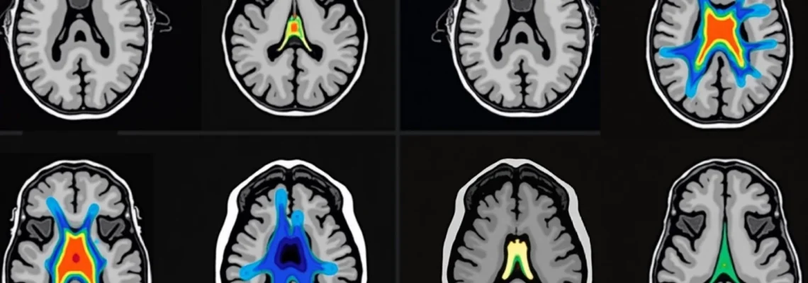
Susceptibility-weighted imaging for central vein sign detection
Susceptibility-weighted imaging (SWI) exploits magnetic susceptibility differences between tissues to enhance visualisation of venous structures and iron deposits. This technique proves invaluable for identifying the central vein sign, a characteristic feature of MS lesions that helps differentiate them from other white matter abnormalities. The central vein sign appears as a small, dark linear structure running through the centre of white matter lesions, reflecting the perivenular distribution of MS pathology.
Research indicates that the central vein sign is present in approximately 80-90% of MS lesions but rarely observed in mimicking conditions such as small vessel disease or migraine-related white matter changes. SWI sequences can identify this diagnostic feature without contrast agents, providing an additional tool for improving diagnostic specificity in challenging cases. The technique requires careful optimisation of acquisition parameters and post-processing algorithms to achieve optimal visualisation of venous structures.
Mcdonald criteria 2017 revisions and Non-Contrast MRI diagnostic standards
The 2017 revision of McDonald criteria marked a significant evolution in MS diagnosis, emphasising the role of non-contrast MRI sequences in establishing both dissemination in space and time. These updated criteria recognise that modern MRI technology can effectively demonstrate the characteristic spatial and temporal patterns of MS pathology without relying on gadolinium enhancement. The revisions specifically acknowledge that new T2 lesions on follow-up scans provide sufficient evidence of dissemination in time, potentially eliminating the need for contrast-enhanced imaging in many clinical scenarios.
A crucial advancement in the 2017 criteria involves the incorporation of cerebrospinal fluid oligoclonal bands as equivalent to dissemination in time when combined with appropriate clinical and imaging findings. This modification enables more rapid diagnosis in patients presenting with clinically isolated syndrome who demonstrate typical MRI lesions but lack temporal dissemination on initial imaging. The criteria maintain strict anatomical location requirements, ensuring diagnostic specificity whilst maximising the utility of non-enhanced sequences.
Dissemination in space requirements using T2-Hyperintense lesions
Spatial dissemination criteria under the 2017 McDonald guidelines require lesions in at least two of four characteristic anatomical regions: periventricular, juxtacortical, infratentorial, and spinal cord. These locations reflect the predilection of MS pathology for specific white matter tracts and regions of high myelin content. T2-hyperintense lesions in these areas demonstrate the multifocal nature of MS, distinguishing it from monophasic conditions such as acute disseminated encephalomyelitis.
The periventricular region requirements focus on lesions adjacent to the lateral ventricles, particularly those demonstrating the characteristic ovoid morphology oriented perpendicular to ventricular surfaces. Juxtacortical lesions must involve the interface between cortex and subcortical white matter, often extending into the cortical ribbon itself. Infratentorial lesions encompass pathology in the brainstem and cerebellum, regions frequently affected in MS and easily identified on non-contrast sequences.
Dissemination in time criteria through serial Non-Enhanced scans
Temporal dissemination traditionally required demonstration of simultaneous enhancing and non-enhancing lesions or development of new lesions on follow-up imaging. The 2017 criteria maintain these principles whilst acknowledging that serial non-enhanced scans can effectively document disease evolution over time. New or unequivocally enlarged T2 lesions on follow-up MRI provide compelling evidence of ongoing disease activity, often eliminating the need for contrast administration in routine monitoring.
Advanced image subtraction techniques significantly enhance the detection of new lesions on serial scans by automatically highlighting areas of interval change. These computational methods subtract baseline images from follow-up scans, effectively cancelling unchanged structures whilst emphasising new pathology. Studies demonstrate that subtraction imaging combined with high-resolution 3D sequences achieves sensitivity approaching that of contrast-enhanced studies for detecting new disease activity.
Juxtacortical and infratentorial lesion location specifications
Juxtacortical lesions represent a distinctive pattern of MS pathology characterised by involvement of both subcortical white matter and overlying cortex. These lesions typically demonstrate a characteristic U-shaped or curved morphology, following the contours of cortical sulci and gyri. The 2017 McDonald criteria specifically recognise cortical lesions as equivalent to juxtacortical lesions for spatial dissemination purposes, reflecting improved understanding of grey matter involvement in MS pathogenesis.
Infratentorial lesions encompass pathology within the brainstem, cerebellum, and their connecting white matter tracts. These locations are readily identified on non-contrast sequences, particularly FLAIR and T2-weighted images, due to excellent contrast resolution in posterior fossa structures. Brainstem lesions often appear as small, focal hyperintensities in characteristic locations such as the medial longitudinal fasciculus or cerebellar peduncles, whilst cerebellar lesions may involve both white matter and cortical regions.
Paramagnetic rim lesions and iron deposition patterns without gadolinium
Paramagnetic rim lesions represent a specific subset of MS pathology characterised by chronic active inflammation and progressive tissue destruction. These lesions demonstrate a distinctive appearance on susceptibility-weighted imaging, with a dark rim of iron deposition surrounding a central core of demyelination. The paramagnetic rim reflects activated microglia and macrophages containing iron deposits, indicating ongoing inflammatory activity even in the absence of blood-brain barrier breakdown. This finding has significant prognostic implications, as paramagnetic rim lesions correlate with more aggressive disease courses and greater disability accumulation.
Detection of paramagnetic rim lesions requires optimised SWI protocols with appropriate echo times and post-processing techniques to enhance susceptibility contrast. Recent studies suggest that approximately 20-30% of MS lesions demonstrate paramagnetic rims, with higher prevalence in progressive disease forms. The ability to identify these lesions without contrast agents provides valuable prognostic information and may influence treatment decisions, particularly regarding the use of more aggressive disease-modifying therapies.
Iron deposition patterns in MS extend beyond individual lesion rims to encompass broader regional changes in susceptibility. Deep grey matter structures, including the thalamus, caudate nucleus, and putamen, frequently demonstrate increased susceptibility on SWI sequences, reflecting both primary pathology and secondary degeneration. These changes correlate with cognitive impairment and physical disability, providing additional biomarkers for disease monitoring without requiring contrast administration. The pattern and extent of iron deposition can help differentiate MS from other neurodegenerative conditions that affect similar brain regions.
Differential diagnosis challenges: MS mimics on unenhanced MRI
Accurate differentiation of MS from mimicking conditions represents one of the most challenging aspects of neuroimaging interpretation, particularly when relying solely on non-contrast sequences. The differential diagnosis encompasses a broad spectrum of conditions that can produce similar-appearing white matter lesions, including vascular, inflammatory, infectious, and genetic disorders. Careful attention to lesion morphology , distribution patterns, and associated clinical features becomes crucial for avoiding misdiagnosis and inappropriate treatment initiation.
The most common MS mimics include cerebral small vessel disease, migraine-related white matter changes, neuromyelitis optica spectrum disorder (NMOSD), and acute disseminated encephalomyelitis (ADEM). Each condition demonstrates characteristic imaging features that can help distinguish it from MS, though overlap exists and may require additional diagnostic testing. Understanding these distinguishing features enables more accurate interpretation of non-contrast MRI studies and reduces the risk of diagnostic errors.
Neuromyelitis optica spectrum disorder lesion characteristics
NMOSD presents distinct imaging characteristics that help differentiate it from conventional MS, particularly on non-contrast sequences. Brain lesions in NMOSD typically occur in characteristic locations including the hypothalamus, brainstem (especially around the fourth ventricle), and areas surrounding cerebral aqueduct. These lesions often demonstrate a different morphology compared to MS, appearing more confluent and less ovoid, with a predilection for midline structures and periependymal regions.
Spinal cord involvement in NMOSD characteristically produces longitudinally extensive lesions spanning three or more vertebral segments, contrasting with the typically shorter lesions seen in MS. These extensive cord lesions often demonstrate central grey matter involvement and may extend from the medulla to the conus medullaris. The combination of characteristic brain lesion distribution and longitudinally extensive spinal cord pathology helps distinguish NMOSD from MS on non-contrast imaging, though serum aquaporin-4 antibody testing remains essential for definitive diagnosis.
Acute disseminated encephalomyelitis pattern recognition
ADEM demonstrates a distinctive imaging pattern that typically differs from MS in several key aspects. The lesions in ADEM are usually more symmetric and bilateral, often involving both grey and white matter structures. Deep grey matter involvement, including the thalamus and basal ganglia, occurs more frequently in ADEM compared to MS, providing an important distinguishing feature. The lesions also tend to be larger and more confluent than typical MS plaques, often demonstrating ill-defined borders.
The temporal evolution of ADEM lesions differs markedly from MS, with most lesions appearing simultaneously and then gradually resolving over weeks to months. This monophasic pattern contrasts with the characteristic temporal dissemination seen in MS, where new lesions continue to develop over time. Serial non-contrast MRI studies can effectively demonstrate this difference in temporal evolution, supporting the correct diagnosis without requiring contrast administration.
Migraine-related white matter hyperintensities distinction
Migraine-associated white matter changes present a common diagnostic challenge, particularly in younger patients with headache disorders. These lesions typically demonstrate a characteristic distribution pattern, predominantly affecting subcortical white matter in frontal and parietal regions whilst sparing the corpus callosum and brainstem. The morphology tends to be more punctate and less ovoid compared to MS lesions, lacking the characteristic perpendicular orientation to ventricular surfaces.
The progression pattern of migraine-related white matter changes differs substantially from MS, typically showing slow accumulation over years rather than the more rapid development characteristic of MS lesions. Additionally, migraine-related changes rarely demonstrate the central vein sign or paramagnetic rim features that are commonly observed in MS lesions on susceptibility-weighted imaging. The absence of spinal cord involvement also helps distinguish migraine-related changes from MS pathology.
Cerebral small vessel disease differentiation techniques
Cerebral small vessel disease represents perhaps the most common MS mimic, particularly in older patients or those with vascular risk factors. The lesion distribution in small vessel disease typically follows vascular territories, with predominant involvement of subcortical white matter and absence of the characteristic periventricular and juxtacortical patterns seen in MS. Lacunar infarcts and microbleeds on susceptibility-weighted imaging provide additional clues to vascular aetiology.
The morphological characteristics of small vessel disease lesions differ from MS plaques in several important ways. Vascular lesions tend to be more rounded and symmetric, lacking the ovoid morphology and Dawson’s finger pattern characteristic of MS. The absence of spinal cord involvement and the association with other markers of cerebrovascular disease, such as cortical infarcts and enlarged perivascular spaces, further support a vascular diagnosis over MS.
Quantitative MRI biomarkers and advanced Non-Contrast techniques
The development of quantitative MRI biomarkers has revolutionised the assessment of MS pathology beyond simple lesion detection, providing insights into tissue microstructure and disease progression mechanisms. These advanced techniques extract quantitative measurements from standard MRI sequences, offering objective metrics for monitoring disease activity and treatment response. Quantitative biomarkers demonstrate particular value in detecting subtle pathological changes that may not be apparent on conventional visual assessment, enabling earlier detection of disease progression and more precise monitoring of therapeutic interventions.
Modern quantitative MRI approaches encompass a diverse range of techniques including magnetisation transfer imaging, diffusion tensor imaging, and volumetric analysis algorithms. These
methods enable comprehensive characterisation of tissue integrity, inflammatory activity, and neurodegeneration processes using conventional MRI hardware without requiring specialised contrast agents or additional scanning time. The integration of these biomarkers into routine clinical protocols provides unprecedented insights into MS pathophysiology whilst maintaining the safety advantages of non-contrast imaging.
Magnetisation transfer ratio measurements in MS pathology
Magnetisation transfer imaging exploits the exchange between mobile protons in free water and immobilised protons bound to macromolecules such as myelin proteins. The magnetisation transfer ratio (MTR) provides a quantitative measure of tissue integrity, with reduced values indicating loss of macromolecular content characteristic of demyelination and axonal damage. This technique demonstrates exceptional sensitivity for detecting subtle myelin loss that may precede the development of visible lesions on conventional sequences, offering potential for earlier disease detection and monitoring.
Clinical studies demonstrate that MTR measurements can differentiate between various types of white matter pathology, with MS lesions typically showing greater MTR reduction compared to age-related changes or small vessel disease. The technique proves particularly valuable for assessing normal-appearing white matter, where significant pathological changes occur despite normal appearance on conventional imaging. Serial MTR measurements enable monitoring of disease progression and treatment response with greater sensitivity than lesion counting alone.
Advanced magnetisation transfer protocols incorporate multiple offset frequencies and flip angles to generate parametric maps of tissue characteristics. These quantitative measurements demonstrate strong correlations with histopathological findings, including myelin density, axonal integrity, and inflammatory cell infiltration. The ability to obtain these measurements from standard pulse sequences makes MTR analysis particularly attractive for routine clinical implementation without additional scanning time or contrast administration.
Diffusion tensor imaging fractional anisotropy analysis
Diffusion tensor imaging (DTI) measures the microscopic motion of water molecules within tissue, providing insights into white matter tract integrity and organisation. Fractional anisotropy (FA) represents the degree of directional preference in water diffusion, with higher values indicating more organised tissue architecture. In MS, demyelination and axonal damage result in decreased FA values, reflecting disrupted tissue microstructure even in areas that appear normal on conventional imaging sequences.
The sensitivity of DTI for detecting MS pathology extends beyond visible lesions to encompass normal-appearing white matter, where significant microstructural changes occur throughout the disease course. Longitudinal DTI studies demonstrate progressive reduction in FA values that correlates strongly with clinical disability measures and cognitive impairment. This technique provides valuable prognostic information and enables monitoring of disease progression with greater precision than conventional lesion-based metrics.
Advanced DTI analysis techniques include tract-based spatial statistics and connectome analysis, which evaluate the integrity of specific white matter pathways and their functional connections. These approaches reveal patterns of tissue damage that may not be apparent from simple FA measurements, providing insights into the mechanisms underlying clinical symptoms and disability progression. The integration of DTI biomarkers into clinical practice offers enhanced capability for personalised treatment monitoring and prognostication.
Brain volumetric assessment using SIENA and SIENAX algorithms
Brain atrophy represents a crucial marker of disease progression in MS, reflecting both grey and white matter volume loss due to inflammatory damage and neurodegeneration. The SIENA (Structural Image Evaluation using Normalisation of Atrophy) algorithm provides automated measurement of brain volume changes between serial scans, whilst SIENAX enables cross-sectional volume measurements normalised for head size. These computational tools achieve exceptional precision in detecting subtle volume changes that would be impossible to assess through visual inspection alone.
Clinical validation studies demonstrate that brain atrophy rates in MS patients significantly exceed normal age-related volume loss, with annual atrophy rates of 0.5-1.0% compared to 0.1-0.3% in healthy controls. The measurement of brain volume loss provides important prognostic information, as higher atrophy rates correlate with more rapid disability progression and cognitive decline. Serial volumetric analysis enables objective monitoring of neuroprotective treatment effects and disease progression independent of inflammatory activity.
Regional volumetric analysis extends beyond global brain measurements to assess specific anatomical structures including cortical thickness, subcortical volumes, and white matter tract integrity. Advanced algorithms such as FreeSurfer and FSL enable detailed parcellation of brain regions, revealing patterns of volume loss that correlate with specific clinical symptoms and functional impairments. The combination of global and regional volumetric measurements provides comprehensive assessment of disease impact across different neural systems without requiring contrast administration.
Clinical limitations and false negative rates in non-enhanced MS imaging
Despite significant advances in non-contrast MRI techniques, certain clinical scenarios present ongoing challenges for accurate MS detection and monitoring. False negative rates, whilst substantially reduced with modern imaging protocols, remain a concern in specific patient populations and disease presentations. Understanding these limitations becomes crucial for clinicians to appropriately interpret non-enhanced studies and determine when additional diagnostic approaches may be necessary. The recognition of these constraints ensures optimal patient care whilst maximising the benefits of contrast-free imaging strategies.
Early disease stages present particular challenges for non-contrast imaging, as initial inflammatory lesions may be small and subtle, potentially escaping detection even with high-resolution sequences. Young patients with mild symptoms may harbour clinically significant pathology that requires careful correlation with clinical findings and potentially serial imaging studies. Additionally, certain anatomical locations, such as the optic nerves and portions of the brainstem, may be difficult to assess adequately without contrast enhancement, particularly when evaluating acute inflammatory activity.
Patient-related factors significantly influence the diagnostic accuracy of non-enhanced MRI studies. Motion artefacts, particularly problematic in patients with tremor or cognitive impairment, can substantially degrade image quality and reduce lesion detection sensitivity. Claustrophobia and inability to remain motionless during lengthy high-resolution acquisitions may necessitate shorter protocols with potentially reduced diagnostic yield. The presence of metallic implants or devices can create susceptibility artefacts that obscure pathology in critical brain regions, limiting the effectiveness of non-contrast techniques.
Technical limitations of non-enhanced imaging include reduced sensitivity for detecting acute inflammatory activity compared to contrast-enhanced studies. While new T2 lesions provide evidence of disease activity, the timing and inflammatory status of lesions cannot be determined as precisely without gadolinium enhancement. This limitation becomes particularly relevant in clinical trial settings where precise measurement of inflammatory activity is required, and in patients with rapidly progressive disease where treatment decisions depend on accurate assessment of current disease activity levels.
The interpretation of non-enhanced studies requires considerable expertise and familiarity with advanced sequence characteristics and normal variants. Radiologists must possess thorough understanding of sequence-specific appearances and potential pitfalls to avoid misinterpretation of normal anatomical structures or benign findings as pathological lesions. The complexity of quantitative biomarker analysis necessitates specialised training and quality assurance protocols to ensure reliable and reproducible measurements across different scanning platforms and institutions.
Future developments in non-contrast MS imaging focus on further improving sensitivity and specificity through artificial intelligence applications and novel sequence development. Machine learning algorithms show promise for automated lesion detection and characterisation, potentially reducing interpretation variability and improving diagnostic accuracy. Advanced techniques such as synthetic contrast imaging and quantitative susceptibility mapping offer potential alternatives to conventional contrast enhancement, providing inflammatory activity information without gadolinium administration. These evolving technologies promise to further expand the capabilities of non-enhanced MRI whilst addressing current limitations and improving patient care outcomes.